

Surgeons who work with millimeter-sized vessels under the microscope: their training, the techniques they use and the conditions they treat
Introduction: A one-millimeter world
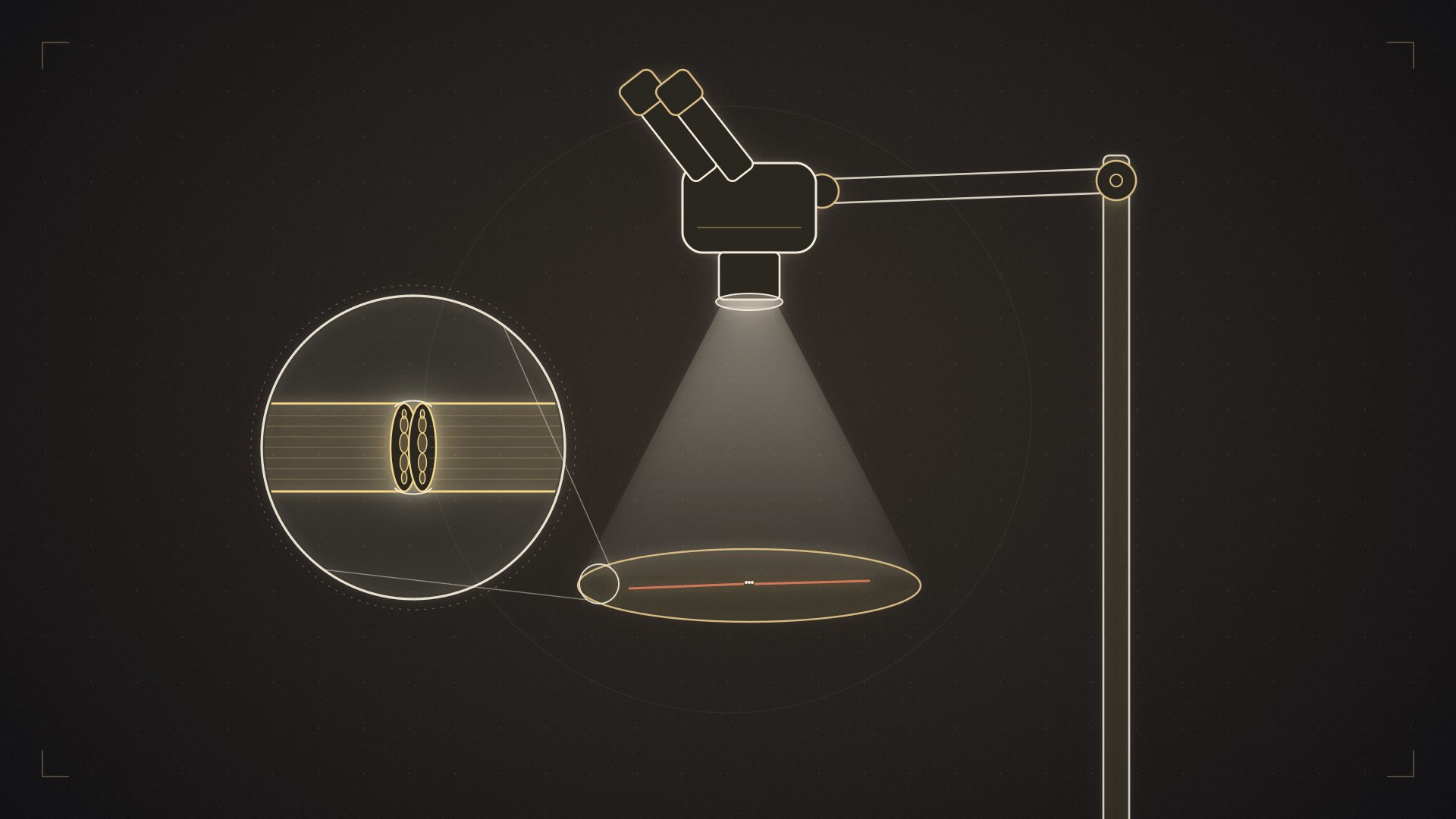
A human hair is about 0.07 millimeters thick. The vessels a microsurgeon routinely sews are 1-3 millimeters in diameter, and the suture thread used is finer than a hair. When working at this scale, even the natural tremor of your hand — everyone has it, and it cannot be prevented — looks like an earthquake under the microscope.
A microsurgeon is a surgeon specially trained to work at this scale. However, it is not a separate specialty: microsurgery is an advanced technique used by more than one specialty. Microsurgeons may come from different fields such as plastic, reconstructive and aesthetic surgery, orthopedics and traumatology, hand surgery, and ear, nose and throat surgery. But being a specialist in one of these fields does not, by itself, mean having advanced microsurgical competence.
This article explains what microsurgery is, how microsurgeons are trained, in which conditions they become involved, and what this means for you as a patient.
If you are looking for a practical guide on how to find the right doctor, that topic is covered in a separate article; this article introduces the field itself.
Important note: This content is for general information only and does not replace examination, diagnosis or treatment by a physician.
1. What is microsurgery?
Definition: A surgical technique in which the surgeon works, under an operating microscope or high-magnification surgical loupes, with structures that cannot be distinguished with the naked eye.
But the real definition is this: microsurgery is the art of moving tissue while keeping it alive.
To understand this, let us make a comparison. When conventional surgery cuts a tissue and places it somewhere else, that tissue loses its own blood supply. Small pieces (such as a skin graft) can survive by being nourished by the bed beneath them. But a thick, bulky or bone-containing tissue cannot survive this way — it dies within a few days.
Microsurgery removed this limit. The tissue is raised together with the artery and vein that nourish it, moved to the target area and sewn to the vessels there under the microscope. Blood flow starts again, and the tissue continues to live in its new location.
The doors this opened:
-
Reattaching a severed finger or arm (replantation)
-
Rebuilding the breast with the patient’s own abdominal tissue
-
Repairing the jawbone with the fibula bone from the leg
-
Restoring a smile to a paralyzed face with a muscle taken from the leg
-
Rebuilding a lost thumb with a toe
-
Connecting blocked lymphatic vessels to veins
None of these is possible without microsurgery.
2. A brief history
Microsurgery is relatively new in the history of medicine.
1921 — An ear surgeon used a microscope in an operation for the first time.
1960s — It was shown that small vessels could be successfully sewn under the microscope. Experimental work accelerated.
1962 — An arm was successfully reattached (replantation) for the first time.
1968 — The first toe-to-thumb transfer: a toe taken from the foot was transplanted to the hand. This was the clinical confirmation of the idea that “tissue can be moved while alive”.
1970s — Free tissue transfer (the free flap) began to spread. It revolutionized reconstruction after cancer surgery and trauma.
1980s-90s — The perforator flap concept developed: it became clear that muscle did not need to be sacrificed to nourish tissue. Techniques such as the DIEP flap are products of this period.
2000s and beyond — Supermicrosurgery was born: working with vessels 0.3-0.8 millimeters in diameter. This made lymphedema surgery possible. In the same period, composite tissue transplants such as hand and face transplants were performed.
Today — Robot-assisted microsurgery systems are being developed, and imaging technologies (ICG fluorescence) are becoming routine.
3. How is a microsurgeon trained?
This is the least known but most decisive aspect of microsurgery.
3.1. The training path
1. Medical school — 6 years.
2. Specialty training — in a main specialty that uses microsurgery: Plastic, Reconstructive and Aesthetic Surgery; Orthopedics and Traumatology; Ear, Nose and Throat; Neurosurgery; Ophthalmology. Typically 4-6 years in Türkiye. Plastic surgery departments have played a pioneering role in the development of microsurgery at university level in Türkiye, and the most comprehensive microsurgical training has traditionally been provided in this field. That said, the scope of training and case volume can vary from center to center, and other specialties can also offer intensive microsurgical experience in their own areas.
3. Subspecialty or advanced training (fellowship) — After specialty training, advanced training focused on microsurgery and reconstruction can be pursued. Fellowship programs, usually lasting 1-2 years, provide advanced clinical experience in surgical planning, delicate tissue dissection, free tissue transfer and complication management at high-volume centers under the supervision of experienced microsurgeons.
There is no official microsurgery subspecialty training in Türkiye. Hand Surgery is a separate subspecialty, and physicians from both plastic, reconstructive and aesthetic surgery and orthopedics and traumatology can pursue it. Although hand surgery training includes important areas that overlap with microsurgery, the title of hand surgery specialist does not, by itself, indicate advanced microsurgical competence. That competence depends on the content of the training, the case variety of the center where the surgeon has worked and the surgeon’s individual microsurgical experience.
4. Laboratory training — this stage cannot be skipped. Basic microsurgical skills are learned not directly on patients but under controlled laboratory conditions. Surgeons develop their technical precision by performing large numbers of vessel repairs, first on silicone tubes, then on chicken wing vessels and, in suitable programs, on experimental animal models. Laboratory training can be obtained in structured courses or at microsurgery training centers, and some programs award a certificate at the end. However, a certificate alone is not an indicator of microsurgical competence. What really matters is that the skill gained in the laboratory is developed through clinical experience and can be applied safely in real operations.
5. Continuous practice — microsurgery is a skill that fades when it is not used. Microsurgery is a skill whose technical precision is maintained through regular practice and which matures with experience accumulated over the years. When it is not practiced for a long time, speed and technical fluency may decline; accumulated experience, on the other hand, provides an important advantage in planning, dissection and making the right decisions, especially in difficult cases. A surgeon who does not operate regularly loses hand precision and speed of decision-making over time.
3.2. Skills that must be learned
Hand control. Everyone’s hand trembles; a microsurgeon cannot eliminate this but learns to manage it: correct posture, supporting the arm and wrist, breath control, the correct way of holding instruments.
Depth perception under the microscope. Working in three dimensions in a field that looks two-dimensional is a separate skill that must be learned.
Vascular repair (anastomosis) techniques. End-to-end, end-to-side, reverse flow — each is used in different situations.
Knowledge of anatomy. Knowing which tissue is supplied by which vessel is the foundation of microsurgery. Mapping out a flap is half the operation.
Tissue dissection. Being able to safely separate vessels and nerves from the surrounding damaged tissue in areas where the anatomy has been distorted by trauma, previous operations, radiotherapy or dense scar tissue is also a microsurgical skill. Tissue dissection, preparing suitable vessels and working without damaging tissue are as important as the vascular repair itself.
Problem-solving. Knowing what to do when a vessel becomes blocked, when lengths do not match or when diameters differ — this is exactly what experience is.
3.3. A microsurgeon’s day
Microsurgical operations are long: 6-12 hours is usual, and some cases take even longer. The surgeon spends a large part of this time at the microscope, motionless and highly concentrated.
Added to this is postoperative follow-up: hourly monitoring of the transferred tissue for the first 48-72 hours and, when necessary, a return to the operating room in the middle of the night. Microsurgery is both physically and mentally demanding, which explains why the number of doctors who choose this field is limited.
4. Tools used in microsurgery
Operating microscope. Provides 6-40x magnification; dual-head models that allow two surgeons to look at the same time are used. Focus and magnification are adjusted with foot pedals.
Surgical loupes (magnifying glasses). 2.5-6x magnification; used for larger vessels and tissue preparation.
Microsurgical instruments. Forceps with tips as fine as a hair, micro-scissors, vascular clamps. These are delicate and expensive instruments.
Suture material. Threads of 9-0, 10-0 and 11-0 gauge. An 11-0 thread is only a few times thicker than a red blood cell; it is almost invisible to the naked eye.
Doppler device. Checks blood flow in the vessel by sound; used during and after surgery. There are also implantable probe models placed on the vessel.
ICG (indocyanine green) fluorescence imaging. A dye given into the vessel shows the tissue’s blood supply in real time with a special camera. It allows the surgeon to see during the operation which part of the flap is well nourished; in lymphedema surgery it is used to map the lymphatic vessels.
Vessel coupling systems. In some venous connections, special coupling rings can be used instead of sutures.
5. What procedures do microsurgeons perform?
5.1. Replantation — reattaching a severed part
Reattaching a completely severed part such as a finger, hand, arm, foot, ear or scalp. Bone fixation, tendon repair, arterial and venous anastomosis, nerve repair and skin closure are performed in sequence. It is a race against time.
5.2. Free tissue transfer (free flap)
Moving tissue taken from one area of the body to another area together with its vessels. It is the most common application of microsurgery. Areas of use: reconstruction after head and neck cancer, breast reconstruction, limb salvage, muscle transfers for function, closure of acute or chronic wounds, bone defects.
At experienced centers, flap survival is usually reported in the range of 95-98%.
5.3. Nerve surgery
Repairing severed nerves, placing nerve grafts in between, nerve transfer (a working nerve taking over the function of a paralyzed muscle). It plays a central role in facial paralysis and brachial plexus injuries.
5.4. Functional muscle transfer
A muscle taken from the leg (usually the gracilis) is transferred with its vessel and nerve and made to work. It is used to restore a smile in facial paralysis, to bring back finger flexion in Volkmann’s contracture, and to provide elbow flexion in brachial plexus injury.
5.5. Toe-to-hand transfer
Rebuilding a lost thumb or fingers with a toe.
5.6. Supermicrosurgery and lymphedema
Connecting lymphatic vessels 0.3-0.8 millimeters in diameter to veins (LVA) and lymph node transfer (VLNT). Used for arm or leg swelling after cancer treatment.
5.7. Composite tissue transplantation
Transplants from another person, such as hand and face transplants. They are performed at a very small number of centers and require lifelong immunosuppressive medication. In this respect they are fundamentally different from other microsurgical procedures that use the body’s own tissue.
6. How does a microsurgical operation work?
Planning. The vessels at the recipient and donor sites are assessed with imaging. Which tissue will be taken, from where and on which vessel — these are decided in advance.
Two teams. In some microsurgical operations, two surgical teams work simultaneously. While one team prepares the recipient site or removes the cancerous tissue, the other prepares and raises the tissue to be transferred. In suitable cases, this way of working can shorten the operation considerably.
Raising the tissue. The flap is dissected while its nourishing vessel is preserved. This stage requires patience and knowledge of anatomy.
Ischemia time. The tissue is without blood from the moment its vessel is cut until it is reconnected. This time is kept as short as possible; tolerance differs by tissue type (muscle is more sensitive than skin).
Anastomosis. Under the microscope, the artery and usually one or two veins are sewn. 8-12 stitches per vessel.
Reperfusion. The clamps are released and blood starts to flow. The tissue regaining color and warmth is the most critical moment of the operation.
Closure and monitoring. The tissue is secured and the skin is closed. Hourly monitoring begins after surgery.
7. The biggest risk: Vascular blockage
This is the risk specific to microsurgery.
What happens? A clot forms in the sewn vessel, blood flow stops and the tissue cannot be nourished. Venous blockage is more common than arterial blockage.
When does it happen? Most cases occur in the first 48-72 hours. This is the reason for intensive postoperative monitoring.
How is it detected? The flap’s color (paling or turning purple), temperature, capillary refill time and Doppler signal are monitored. Observation by experienced nurses is decisive here.
What is done? The patient is taken urgently to the operating room, and the clot is removed or the anastomosis is redone. With early intervention, the chances of saving the tissue are high; delay lowers these chances rapidly.
That is why: At a center that performs free tissue transfer, having 24-hour emergency surgical capability is not a technical detail but a fundamental safety requirement.
8. Factors that affect microsurgical success
Surgeon-related: Experience, case volume, choosing the right flap, technical meticulousness, keeping ischemia time short.
Center-related: Two-team capacity, quality of monitoring, 24-hour emergency capability, anesthesia experience, rehabilitation support.
Patient-related:
-
Smoking — one of the most decisive factors; nicotine constricts the vessels and directly increases the risk of blockage
-
Vascular disease, advanced diabetes, advanced age
-
Previous radiotherapy to the area (makes the vessels more difficult)
-
Nutritional status and blood values
-
Clotting disorders
-
Compliance with postoperative instructions
Injury-related: In crush-type injuries the vessels are damaged over a wide area, and the success rate drops. Results are better with clean cuts.
9. What does microsurgery mean in different specialties?
| Specialty | What it does with microsurgery |
|---|---|
| Plastic and Reconstructive Surgery | Free flaps, breast reconstruction, replantation, facial paralysis, lymphedema, complex wounds, toe-to-hand transfer, nerve repair, brachial plexus, etc. (any area of microsurgery, depending on the surgeon’s interests) |
| Hand Surgery (subspecialty) | Replantation, toe-to-hand transfer, nerve repair, brachial plexus |
| ENT / Head and Neck Surgery | Surgical treatment of head and neck tumors; multidisciplinary planning and repair, together with Plastic Surgery, of the resulting tongue, jaw, palate and neck defects. Those with microsurgical training can take part in reconstructive procedures. |
| Neurosurgery | Aneurysm clipping, bypass, peripheral nerve repair |
| Ophthalmology | Retinal and anterior segment microsurgery (a different branch of microsurgery) |
| Cardiovascular Surgery | Small vessel repairs, vascular bypass |
| Urology / Gynecology | Reconstructive procedures, microsurgical varicocele repair, fallopian tube repair |
10. What does this mean for you as a patient?
One: The name of your surgeon’s specialty does not, by itself, tell you whether they perform microsurgery. What you need to ask about is that surgeon’s experience with this procedure.
Two: Microsurgical operations take a long time and require intensive monitoring. This is why the length of hospital stay, postoperative restrictions and the smoking ban are strict — not arbitrary, but a biological necessity.
Three: Success rates are high but not 100%. There is always a chance of tissue loss, and this is not necessarily the surgeon’s fault.
Four: Flap survival is not the end of the operation but the beginning. The real measure is function: can the hand grip, can the face smile, has the patient returned to work? Rehabilitation is essential for this.
Five: These operations are often staged. Secondary revision procedures (thinning, scar revision, tendon release) are normal and do not mean failure.
11. Donor sites commonly used in microsurgery
One of a microsurgeon’s most important areas of knowledge is knowing which tissue can be taken from which part of the body, and on which vessel. This map is called flap anatomy, and it forms the backbone of the field.
Commonly used donor sites:
| Donor site | What it provides | Typical use |
|---|---|---|
| Anterolateral thigh (ALT) | Large area of skin and soft tissue, long vascular pedicle | Head and neck, limbs, abdominal wall — the main “workhorse” flap, the most widely used |
| Lower abdomen (DIEP) | Skin and fat; the muscle is preserved | Breast reconstruction |
| Outer bone of the lower leg (fibula) | Bone and, if needed, skin | Jaw and long-bone reconstruction |
| Back muscle (latissimus dorsi) | Large muscle and skin | Extensive tissue loss, functional muscle transfer |
| Inner thigh muscle (gracilis) | Slender muscle that can be reinnervated | Facial paralysis, Volkmann’s contracture, brachial plexus |
| Forearm (radial forearm) | Thin, pliable skin; long vascular pedicle | Reconstruction of the inside of the mouth, the tongue, the penis |
| Shoulder blade region (scapula) | Bone and skin together | Complex facial reconstruction |
| Toe | A jointed toe with a nail and sensation | Finger reconstruction |
| Groin / intra-abdominal lymph nodes | Lymphatic tissue | Lymphedema surgery |
What is the choice based on? The type of tissue lost (skin, bone or muscle), the volume needed, the length and diameter of the vascular pedicle, the scar and loss of function left at the donor site, and the patient’s build and previous operations.
The “replace like with like” principle. This is the fundamental rule of reconstructive surgery: thin skin is brought where thin skin is needed, bone where bone is needed, innervated tissue where sensate tissue is needed. That is why thin forearm skin is moved into the mouth, fibula bone to the jaw, and a muscle that can move to the face.
12. Differences between microsurgery and conventional surgery
| Conventional surgery | Microsurgery | |
|---|---|---|
| Vision | Naked eye | Microscope or loupes (6-40x) |
| Structures worked on | Larger than a few millimeters | 0.3-3 millimeters |
| Sutures | Visible to the eye | Finer than a hair (9-0 to 11-0) |
| Duration | Usually short to medium | 6-12 hours is usual |
| Team | A single team may be enough | Two teams not mandatory but preferred |
| After surgery | Standard follow-up | Hourly vascular monitoring for the first 72 hours |
| Main risk | Bleeding, infection | In addition to these, vascular blockage |
| Learning | In the operating room | First in the laboratory, then in the operating room |
The practical meaning of these differences: Extensive microsurgical operations require special organization on the part of the hospital. Long operating-room time, a trained team, close monitoring, intensive care support when needed and the capacity for urgent re-operation are important. The surgeon’s long training, the high cost of the operations and the hospital infrastructure required explain why the number of surgeons and centers working in this field is limited.
13. The other members of the team
Microsurgery looks like one surgeon’s work, but it is not.
Anesthesia team. In long operations, maintaining the patient’s temperature, blood pressure and fluid balance is critical. A drop in body temperature constricts the vessels and increases the risk of blockage; keeping the patient warm is the anesthesia team’s job. Blood pressure must be kept neither too high nor too low — both threaten the flap.
Operating room nurse. Microsurgical instruments are delicate and must be passed in the right order and in the right way. An experienced microsurgical nurse shortens the operation considerably.
Ward and intensive care nurses. They are the people who actually carry out flap monitoring. The nurse is usually the first to notice a change in color, temperature or capillary refill, and that difference determines whether the tissue can be saved.
Physiotherapist / hand therapist. In functional operations, they are the ones who determine the result. After a nerve transfer or muscle transfer, the brain learns the new connection through months of work with a therapist.
Radiology. Preoperative vascular mapping (CT angiography, Doppler) and, when needed, postoperative assessment.
Pathology and oncology. Joint work on margin safety and the treatment plan in reconstructions performed together with cancer surgery.
Psychological support. In patients going through limb loss, facial change or a long rehabilitation process, it directly affects adherence to treatment.
14. The future of microsurgery
Robot-assisted microsurgery. Robotic systems that filter hand tremor and scale movement hold promise, especially in supermicrosurgery. They are not yet a widespread standard.
Advanced imaging. The spread of ICG fluorescence imaging allows the tissue’s blood supply to be assessed objectively during surgery.
Tissue engineering. Laboratory-produced tissue scaffolds and bioprinting technologies may reduce the need for donor sites in the long term. They are not yet part of clinical routine.
Immunology in composite tissue transplantation. Protocols that would reduce the need for immunosuppressive medication in hand and face transplants are being researched.
Simulation and training. Virtual reality-based microsurgery training is becoming a tool that supports laboratory training.
15. Frequently asked questions
Is microsurgery a separate specialty?
No. It is an advanced technique used by more than one specialty. In Türkiye, Hand Surgery is a separate subspecialty, but it does not guarantee microsurgical training.
Does every plastic surgeon perform microsurgery?
No. Microsurgery is taught during plastic surgery specialty training, but the number of surgeons who regularly perform free flaps is limited. What you need to ask about is the surgeon’s case volume. You can ask your plastic surgeon directly whether they perform microsurgery; an experienced surgeon will usually give a clear answer.
Why do microsurgical operations take so long?
Sewing vessels under the microscope is slow work. When tissue preparation, transfer, bone fixation and tendon and nerve repair are added, 6-12 hours is usual.
Is it normal to have two surgeons in the operation?
Yes, it is the preferred situation. When two teams work in parallel, the operation is shorter and the tissue spends less time in ischemia (without oxygen).
Why is smoking so important?
Nicotine constricts the vessels. In microsurgery, the vessels connected are 1-3 millimeters in diameter; constriction directly means blockage. Nicotine patches and e-cigarettes have the same effect.
Is microsurgery the same as robotic surgery?
No. Microsurgery is performed by hand under the microscope. Robot-assisted microsurgery systems are being developed but are not yet widespread.
If the vessel becomes blocked, is the tissue definitely lost?
No. If it is noticed early and treated urgently, the chances of saving it are high. That is why close monitoring is carried out in the first 72 hours.
Will the transferred tissue have sensation?
It depends. If a nerve repair was performed, sensation returns over time, but it may not fully reach normal levels. With some flaps sensation remains limited, while with others a certain level of sensation returns even without nerve repair.
Does the transferred tissue grow?
In children, yes — a transferred toe or bone grows with the child. In adults, the tissue continues to behave like the area it was taken from (for example, tissue taken from the abdomen can grow with weight gain).
Can I have an MRI after microsurgery?
Usually yes; it depends on the fixation material used. Ask your surgeon and keep your implant information.
What is the standard of microsurgery in Türkiye?
Microsurgery in Türkiye has a well-established tradition; replantation and free tissue transfer are performed routinely in university hospitals and large training and research hospitals.
Summary: 8 things to remember
-
Microsurgery is not an independent specialty but an advanced surgical technique used in different surgical fields.
-
Its basic purpose is to precisely repair very small vessels and nerves; transferring tissue with its own vessels to another area is one of its important applications.
-
Microsurgical skill begins with laboratory training and develops through supervised operative experience; it is maintained through accumulated experience and regular practice.
-
Vessels of 1-3 millimeters are routine; in supermicrosurgery, 0.3-0.8 millimeters.
-
The biggest risk is vascular blockage, which mostly develops in the first 48-72 hours.
-
24-hour emergency surgical capability is indispensable for a center that performs microsurgery.
-
Smoking is the patient-related factor that most affects success.
-
Flap survival is not enough — the real measure is the function gained, and that comes with rehabilitation.
To see patients’ questions about preoperative preparation, hospital stay, risks, recovery and the different reconstruction options in one place, read our guide 35 Most Frequently Asked Questions About Microsurgical Reconstruction.
For a personal assessment of your situation, you can request an evaluation appointment; to learn about the surgeon’s training and experience, you can review the surgeon’s profile.
This content has been prepared for general information purposes and does not replace examination, diagnosis or treatment by a physician.
Assoc. Prof. Ahmet Hamdi Sakarya, MD, MSc
Specialist in Plastic, Reconstructive and Aesthetic Surgery
Assoc. Prof. Ahmet Hamdi Sakarya completed a two-year fellowship in reconstructive microsurgery and peripheral nerve surgery at Chang Gung Memorial Hospital in Taiwan, one of the world’s leading high-volume microsurgery centers. His clinical practice focuses on facial paralysis and facial reanimation, brachial plexus and other peripheral nerve injuries, functional muscle transfers, free tissue transfers and facial aesthetic surgery.


