

Moving tissue from one part of the body to a distant area: the flaps used, success rates, vascular monitoring and recovery
Introduction: The end of the distance limit
For a long time, the biggest constraint of reconstructive surgery was this: tissue could only be moved as far as its vessel could reach. A surgeon who wanted to bring abdominal skin to the leg had only one option — keeping the patient’s arm stitched to the abdomen for weeks.
In the 1970s, the development of microsurgery removed this limit. Tissue could now be taken by dividing its nourishing vessel, moved anywhere in the body and sewn to new vessels there under the microscope.
This is called a free flap — or free tissue transfer — and today it is the most powerful tool in reconstructive surgery. Rebuilding the jawbone with bone taken from the leg, recreating the breast with the patient’s own abdominal tissue, saving a crushed leg — all of these are possible with this technique.
This guide explains free flap surgery from a patient’s perspective: how it is done, which tissue is taken from where, what the success rates are, what happens in the first 72 hours and how recovery really works.
If you are curious about the general framework of flap surgery (local flaps, the difference from grafts, classifications), that topic is covered in a separate guide; this article focuses only on free flaps.
Important note: This content is for general information only and does not replace examination, diagnosis or treatment by a physician.
1. What is a free flap?
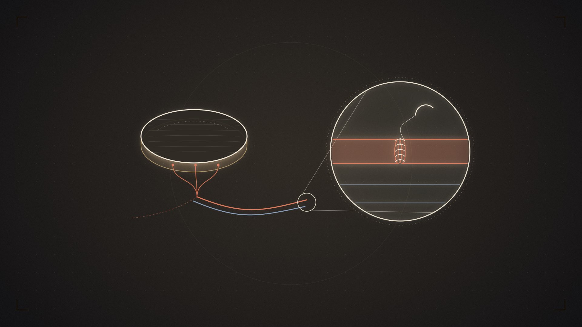
Definition: A piece of tissue that is completely detached (freed) from one part of the body together with its nourishing artery and veins, moved to a distant area and connected to the vessels there under the microscope.
What does “free” mean? The tissue is severed from all its connections to the site where it was taken — it is literally free. This means it is left without blood (ischemic) for anywhere from a few minutes to a few hours. Once its vessels are connected at the new site, blood flow returns and the tissue continues to live.
The difference from a pedicled flap: In a pedicled flap, the tissue is rotated while remaining attached to its vessel; the distance it can reach is limited by the length of the vascular pedicle. A free flap has no such limit.
What can be transferred? Skin, fat, fascia, muscle, vascularized nerve, bone, a segment of bowel, omentum (the fatty apron inside the abdomen), lymph nodes, a toe — and combinations of these.
2. How is the operation performed?
1. Planning. The defect is assessed, and the type and amount of tissue needed are determined. Vascular mapping is performed: both the location of the perforator vessels at the donor site and the suitability of the vessels to be connected at the recipient site are examined with Doppler ultrasound or CT angiography and marked on the skin.
2. Two teams. If two experienced surgical teams are available, they work in parallel: one prepares the recipient site (removing dead tissue, finding the vessels) while the other raises the flap. This significantly shortens the operation and reduces the tissue’s ischemia time.
3. Raising the flap. The tissue is dissected (carefully freed from the surrounding tissues) while its nourishing vessel is preserved. In perforator flaps, this stage requires separating the vessel from the muscle millimeter by millimeter and can take hours.
4. Dividing the vessel. The flap is now free. The ischemia time — without oxygen or blood — begins. This period is kept as short as possible; flaps containing muscle tolerate ischemia less well than skin flaps.
5. Inset and fixation. The flap is placed in the defect; if it contains bone, it is fixed with plates and screws.
6. Vascular repair (anastomosis). The heart of the operation. Under the microscope, one artery and usually one or two veins are sewn. Each vessel takes 8-12 stitches, each with a thread finer than a human hair.
7. Restoring blood flow (reperfusion). The clamps are released and blood starts to flow. The flap turning pink and warm is the most critical moment of the operation. If there is a problem, this is the point at which its cause is carefully sought. It is the most critical moment for identifying and solving any problem.
8. Closure. The skin is closed and a drain is placed. If necessary, a monitoring window is left over the flap.
9. Closing the donor site. It can be closed directly or may require a skin graft.
Duration: Usually 6-12 hours; longer in complex cases.
3. Commonly used free flaps
| Flap | Where it is taken from | What it provides | Typical use |
|---|---|---|---|
| ALT (anterolateral thigh) | Front-outer surface of the thigh | Large area of skin and fat; long vascular pedicle | Head and neck, limbs, abdominal wall — the most widely used flap |
| DIEP | Lower abdomen | Skin and fat; the muscle is preserved | Breast reconstruction |
| Fibula | Outer bone of the lower leg | Long bone; skin if desired | Jaw reconstruction, long-bone defects |
| Radial forearm | Inner surface of the forearm | Very thin, pliable skin; long vessel | Reconstruction of the inside of the mouth, the tongue, the penis |
| Latissimus dorsi | Back | Large muscle and skin | Extensive tissue loss, breast, limbs |
| Gracilis | Inner thigh | Slender muscle that can be reinnervated | Facial paralysis, functional muscle transfer |
| Scapular/parascapular | Shoulder blade region | Bone and skin together | Complex facial reconstruction |
| Free toe | Foot | A jointed toe with sensation | Finger reconstruction |
| Omentum | Inside the abdomen | Well-vascularized, moldable tissue | Infected defects, lymphedema |
| Jejunum | Small intestine | Tube-shaped tissue | Esophageal reconstruction |
The ALT flap — why is it the main “workhorse” flap?
The ALT (anterolateral thigh) flap, taken from the front-outer surface of the thigh, is the most widely used option in modern reconstruction. The reasons:
-
A long vascular pedicle (8-16 cm) and adequate vessel diameter (2-2.5 mm) — ideal for microsurgery
-
A large area of skin can be taken; the donor site can usually be closed directly
-
Adaptable: it can be thinned, taken together with muscle, or divided into several parts
-
Low donor-site morbidity — thigh function is preserved
-
It can be harvested without turning the patient face down, in the same position
Success rates of over 95% have been reported in the literature for the ALT flap.
The DIEP flap — the standard for breast reconstruction
Skin and fat from the lower abdomen are taken while preserving the abdominal muscle. The perforator vessel is dissected out through the muscle.
Advantage: A natural look and feel with the patient’s own tissue; no implant is needed; because the abdominal muscle stays in place, the risk of hernia and abdominal weakness is significantly lower than with older methods that took the muscle. In addition, the abdomen becomes flatter — a kind of tummy tuck at the same time.
Limitation: It may not be suitable for slim patients who do not have enough abdominal tissue; planning is more difficult in patients who have had previous abdominal surgery.
The fibula flap — the standard for jaw reconstruction
The outer bone of the lower leg (the fibula) is taken, together with the overlying skin when needed. It is cut and shaped to recreate the shape of the jaw and fixed with plates. Dental implants can be placed in it. It is frequently used after head and neck cancers, tumors and accidents.
Donor site: The main weight-bearing bone of the leg (the tibia) stays in place; walking is preserved. A segment is left at the lower end to keep the ankle stable.
4. When is a free flap needed?
After head and neck cancer. Repair after removal of tissues of the tongue, floor of the mouth, jaw, palate, cheek and neck. The goal is to regain speech and swallowing function.
Breast reconstruction. Rebuilding the breast with the patient’s own tissue after mastectomy.
Limb salvage. In severe leg and arm injuries where bone and tendon are exposed, to prevent amputation. In open fractures of the lower leg, a free flap is often mandatory.
Bone defects. Vascularized bone transfer after tumors, after infection or in fractures that fail to heal.
Chronic and infected wounds. Bone infection (osteomyelitis), wounds that do not heal after radiotherapy, exposed implants.
Functional reconstruction. Muscle transfer for smiling in facial paralysis; finger flexion in Volkmann’s contracture; elbow flexion in brachial plexus injuries.
Finger and hand reconstruction. Toe-to-hand transfer.
Lymphedema surgery. Vascularized lymph node transfer (VLNT).
Reconstruction of the esophagus and abdominal wall.
5. Success rates: The numbers
Overall free flap survival: In experienced hands it is usually reported in the range of 95-98%.
ALT flap: Success above 95% in the literature.
In difficult patient groups: In challenging scenarios such as high-energy trauma, the rates drop. A systematic review examining the use of the ALT flap in lower-limb trauma found a flap survival rate of 92.4%. The same review reported that the complication rate in high-energy trauma patients was markedly higher than the literature average, but that a high rate of limb salvage was achieved — in other words, partial complications did not mean amputation.
How should these numbers be read?
-
Free flap surgery is a reliable field
-
However, the chance of failure is not zero — a claim of “100% success” is not consistent with the literature
-
The rates depend on the experience of the surgical team; the same results should not be expected at low-volume centers
-
The patient profile is decisive: planned cancer surgery and a crush injury are not the same group
And most importantly: Flap survival is necessary for a successful operation, but not always sufficient. The more valuable measure is function — can the patient speak, swallow, walk, return to work? However, return of function may not be possible for every patient.
6. The most critical period: The first 72 hours
The specific risk of free flap surgery is clot formation at the anastomosis and cessation of blood flow.
When does it happen? The vast majority of cases occur in the first 48-72 hours.
Which is more common? Venous blockage is more common than arterial blockage.
How is the flap monitored?
| Finding | Normal | Arterial problem | Venous problem |
|---|---|---|---|
| Color | Pink | Pale, white | Purple, dark |
| Temperature | Warm | Cold | Warm at first, then cold |
| Capillary refill | 2-3 seconds | Slow or absent | Very fast (under 1 second) |
| Pinprick test | Bright red | No bleeding | Dark blood, flows quickly |
| Tension | Soft | Flaccid | Tense, swollen |
Methods used: Clinical observation (the most basic and most valuable method), handheld Doppler, implantable Doppler probe, tissue oxygen measurement, ICG fluorescence imaging.
Frequency: Usually hourly for the first 24-48 hours, then continued at decreasing intervals.
What happens if a problem is noticed? The patient is taken urgently to the operating room; the clot is removed or the anastomosis is redone. With early intervention, the chances of saving the flap are significantly higher; every hour that passes lowers those chances.
That is why: At a center that performs free flaps, 24-hour emergency surgical capability — with its surgeon, anesthesia team and operating room — is not a technical detail but a fundamental safety requirement. Be sure to ask this question when choosing a center.
7. Risks and complications
Total flap loss. The most serious complication; its rate is low at experienced centers but not zero. If it happens, a new reconstruction is planned.
Partial flap loss. More common; usually managed with dressings, debridement or a small additional procedure.
Vascular crisis and return to surgery. Described above.
Hematoma and seroma. They can compress the flap and disrupt its circulation; drainage may be needed.
Infection. Especially in contaminated and chronic wounds.
Wound dehiscence, fistula. A salivary fistula can occur after reconstructions of the inside of the mouth and the esophagus.
Donor site problems. Wound-healing problems, seroma, scarring, altered sensation, reduced muscle strength, risk of hernia with abdominal flaps, temporary ankle discomfort after fibula harvest.
Aesthetic issues. The flap may differ in color and thickness from the surrounding tissue and give a “patch” appearance. This is corrected with secondary thinning procedures.
General risks. Long anesthesia, blood loss and the need for transfusion, deep vein thrombosis, lung complications, pressure sores (related to the long operating position).
Factors that increase the risk: Smoking (the most decisive), vascular disease, uncontrolled diabetes, an irradiated recipient site, poor nutrition, advanced age, obesity, clotting disorders, crush-type injuries.
8. Preparing for surgery
Smoking. This is the strictest rule for this operation. Nicotine constricts vessels just 1-3 millimeters in diameter and directly creates a risk of blockage. Most centers require patients to quit at least 4-6 weeks beforehand and not to smoke for the same period after surgery. Nicotine patches and e-cigarettes have the same effect.
Vascular assessment. A healthy vessel that can be connected must be available at the recipient site. This assessment is critical in leg injuries and in irradiated areas.
Nutrition. In cancer patients and patients with chronic wounds, nutritional support directly affects healing.
Blood sugar and blood pressure control.
Medication adjustment. Blood thinners and supplements that increase bleeding are managed under a physician’s supervision.
Dental and infection sources. Especially in head and neck reconstructions, a preoperative dental assessment is standard.
Expectations discussion. How many stages the process will involve, that the appearance in the first months will not represent the final result, and that revision procedures are normal should all be discussed in advance.
9. After surgery: The real process
| Period | What happens |
|---|---|
| 0-72 hours | The most critical period. Hourly flap monitoring, positioning restrictions, the room is kept warm, smoking and caffeine forbidden, balanced blood pressure |
| Days 3-7 | Circulation stabilizes. Drains are removed. Mobilization begins |
| Weeks 1-2 | Stitch removal, discharge (usually after 5-14 days). The flap looks swollen and thick |
| Weeks 2-6 | Swelling decreases, activity increases, the donor site heals. A radiotherapy plan begins if needed |
| Weeks 6-12 | The tissue softens, physiotherapy intensifies. Swallowing and speech therapy for head and neck patients |
| Months 3-6 | Swelling largely subsides; sensation begins to return partially |
| Months 6-12 | The tissue matures; the final appearance becomes clear |
| Months 12-24 | Revision procedures: thinning, contour correction, scar revision, dental implants |
Critical rules (first weeks):
-
No pressure is applied to the area of the flap — do not lie on it, and avoid tight clothing and tight bandages
-
The skin portion of the flap is not fully covered, so that any color changes can be observed
-
Keep it warm — getting cold constricts the vessels
-
No smoking
-
Plenty of fluids; blood pressure should be neither too low nor too high
-
With lower-leg flaps, the leg is elevated and then gradually switched to dangling
Report immediately: Any change in the flap’s color, suddenly increasing pain, fever, discharge or a bad smell, sudden swelling.
10. A framework for expectations
Realistic goals:
-
Closing the defect with living tissue and healing the wound
-
Saving the limb or organ
-
Regaining function (speech, swallowing, walking, gripping) — not possible for every patient
-
Bringing infection under control
-
An acceptable appearance and a return to social life
Unrealistic goals:
-
An exact return to the preoperative appearance
-
A process that is completed in a single operation
-
A perfect color and texture match between the transferred tissue and its surroundings
-
Completely normal sensation
Secondary procedures are normal. In free flap reconstructions, thinning, contour correction, scar revision and functional corrections are a planned part of the process — not a sign of failure. Thinking of the process as staged is the most realistic approach.
11. Challenging scenarios
Some situations in free flap surgery are significantly more difficult, and knowing this matters for managing expectations.
Irradiated areas. Radiation makes vessels fragile, impairs tissue healing and increases scar tissue. Finding recipient vessels becomes harder; sometimes vessels are sought outside the radiation field (for example, if there is no suitable vessel in the neck, extension grafts from the chest region can be used). In these patients, a flap that brings its own circulation rather than a graft is mandatory anyway.
Crush-type injuries. In high-energy trauma, the vessels are damaged over a wide area — a vessel that looks intact from the outside may be injured on the inside. This requires extending the dissection beyond the zone of injury so that the anastomosis can be made to healthy tissue.
The lower leg. One of the most challenging reconstruction areas in the body: the bone lies very close to the skin, circulation is limited, swelling is common and gravity works against the flap. Despite this, limb salvage rates are high.
Patients with diabetes and vascular disease. The quality of the recipient vessels is decisive; when necessary, a vascular intervention (balloon, stent, bypass) can be performed first.
Repeat reconstructions. If a previous flap failed, the recipient vessels may already have been used. In that case, more distant vessels or vein extension grafts come into play.
Obesity. It creates additional challenges in terms of flap thickness, donor-site healing and anesthesia.
What changes for these patients? Success rates drop somewhat, complication rates rise and the process usually proceeds in more stages. But “difficult” does not mean “impossible” — in experienced hands, high rates of limb and organ salvage are reported in this group as well.
12. Rehabilitation and functional outcome
Flap survival is not the end of the process but the beginning. The functional outcome is determined by rehabilitation, which requires different teams depending on the area.
In head and neck reconstruction: Swallowing therapy, speech therapy, mouth-opening exercises and, if needed, dental rehabilitation and implant planning. Returning to intelligible speech after tongue reconstruction is directly linked to therapy.
In limb reconstruction: Preserving joint range of motion, gradual weight-bearing, gait training, edema management and, when needed, orthoses and special footwear. For lower-leg flaps, gradual dangling training (dependency training) is a specific program: the leg is lowered little by little over days to weeks so that the flap can adapt to this position.
In hand reconstruction: Tendon gliding, sensory re-education and grip exercises with a hand therapist.
In functional muscle transfers: Biofeedback and mirror exercises so that the brain can learn the new connection; this process takes months.
In breast reconstruction: Shoulder range of motion, scar massage and, if necessary, lymphedema follow-up.
Psychosocial support. In the reconstruction of visible areas such as the face and hands, body image and the process of returning to social life should not be neglected. This support is especially valuable for patients after cancer surgery.
Key point: When choosing a center, the question “who will manage rehabilitation?” is as decisive as “which flap will you use?”
13. Choosing a center
Free flap surgery is an operation that cannot be performed at every hospital or by every surgeon. What to look for:
-
A surgeon with extensive microsurgical experience and, if possible, two-team capacity
-
24-hour emergency surgical capability — this is non-negotiable
-
Experienced nurses or resident doctors trained in flap monitoring
-
An intensive care / close-monitoring bed (not needed for every flap, but the infrastructure must be ready)
-
An anesthesia team experienced in long operations
-
Rehabilitation support (a speech and swallowing therapist for head and neck cases, a physiotherapist for limbs) (they do not have to be at the same center)
-
A multidisciplinary structure working with oncology (for cancer cases)
Questions to ask:
-
What is your annual and total free flap experience? What is your flap loss rate?
-
Which flap do you choose and why? What are the alternatives?
-
What will I lose at the donor site?
-
Who will monitor the flap after surgery, and how often?
-
What happens if there is a vascular problem at midnight — can emergency surgery be performed?
-
How many stages will the process involve?
-
Who will manage rehabilitation?
-
If I am going to receive radiotherapy, how will the timing be planned?
Warning signs: “100% success” or a guaranteed result, a claim of “I have never lost a flap”, no clear answer to the question about emergency surgical capability, glossing over the donor site, being told it will all be done in one operation.
14. My personal approach to free flap surgery
During my two-year reconstructive microsurgery fellowship at Chang Gung Memorial Hospital in Taiwan and my subsequent years as a faculty member, I performed a wide variety of free flap operations. The options in which I have the most experience in my own practice include the ALT flap, the fibula flap and the gracilis muscle flap. I use the ALT flap to repair soft-tissue defects in different parts of the body, particularly the head and neck; the fibula flap especially for jaw reconstruction; and the gracilis muscle flap to restore smiling in facial paralysis or lost movement of the arm and elbow.
In my approach, I care not only about the survival of the transferred tissue but also about minimizing, as far as possible, the functional and aesthetic loss at the site from which the tissue is taken. Especially with ALT and fibula flaps, I prioritize dissection techniques that preserve muscles, nerves and important surrounding structures. In this way, my aim is to avoid creating unnecessary loss in the patient’s healthy area while carrying out the reconstruction.
For microvascular connections, I use the interrupted-continuous vessel suturing technique, which I learned during my training in Taiwan, later adapted to my own practice, and which our team developed further and published as a contribution to the literature. This approach helps make vascular repair controlled, standardized and reliable. In jaw reconstructions, in suitable patients, I aim to restore sensation to the lower lip by including the sensory nerve in the repair.
Free flap surgery is a very broad field, and reconstruction of each area requires different technical knowledge and experience. For this reason, I choose the flap not only according to the size of the tissue defect, but by jointly considering the function needed, the structure of the tissue, preservation of the donor site and the patient’s long-term quality of life.
15. Frequently asked questions
What is the difference between a free flap and a pedicled flap?
In a pedicled flap, the tissue remains attached to its vessel and is moved a limited distance. In a free flap, the vessel is divided, the tissue is moved to a distant area and sewn to new vessels under the microscope.
Why does the operation take so long?
Dissecting, transferring and fixing the flap, and preparing and suturing the vessels under the microscope, all take time. 6-12 hours is usual.
Does the flap have sensation?
Usually limited. Sensation is better if a nerve is also transferred. Over time some sensation returns from the surroundings, but it may not reach normal levels.
Does the transferred tissue grow when I gain weight?
Yes, it continues to behave like the area it was taken from. A flap taken from the abdomen can grow with weight gain; this is taken into account in breast reconstruction.
Will hair grow on the flap?
It depends on where it was taken from. Forearm skin transferred inside the mouth may grow hair; this is considered during planning, and hair removal is performed if necessary.
I am going to receive radiotherapy — will the flap be affected?
Radiotherapy can affect the flap (shrinkage, hardening). The timing is planned together with the oncology team. This is an important topic to discuss before surgery.
What happens if the flap is lost?
A new reconstruction is planned — a second free flap, a pedicled flap or a different method. It is rare, but it can happen.
How long will I stay in hospital?
Usually 5-14 days. The main factor determining the length of stay is flap monitoring in the first days.
When can I return to work?
It varies with the procedure, the location of the flap and your occupation: from a few weeks to a few months.
Is smoking really that important?
Yes. It is the strictest rule for this operation, and most centers require patients to quit.
Is free flap surgery covered by the Turkish Social Security Institution (SGK)?
In state hospitals, it is generally covered for reconstructive indications; coverage varies by procedure and institution. For private hospitals, ask about the terms of your private insurance.
Can I come from abroad or from another city for surgery?
It is possible, but because the first 72 hours are critical and emergency surgery may be needed if a complication occurs, a realistic length of stay is 2-4 weeks. It may be longer depending on the area and extent of the surgery. Book a flexible return ticket and plan in advance how your rehabilitation will continue in your own country or city.
Summary: 8 things to remember
-
In a free flap, the tissue is moved by dividing its vessel and is connected to a vessel at its new site under the microscope — there is no distance limit.
-
The ALT flap is the most widely used option; DIEP is the standard for the breast and the fibula for the jaw.
-
The perforator technique made it possible to take skin without sacrificing muscle — significantly reducing donor-site loss.
-
The success rate at experienced centers is 95-98%, lower in difficult trauma groups. A “100%” claim is not consistent with the literature.
-
The first 48-72 hours are critical. Venous blockage is more common than arterial blockage.
-
A pale, cold flap suggests an arterial problem; a purple, tense flap suggests a venous problem — both are emergencies.
-
A free flap at a center without 24-hour emergency surgical capability is an unnecessary risk.
-
Secondary revision procedures are part of the plan — a flap is not judged by how it looks in the first months.
For a personal assessment of your situation, you can request an evaluation appointment; to learn about the surgeon’s training and experience, you can review the surgeon’s profile.
This content has been prepared for general information purposes and does not replace examination, diagnosis or treatment by a physician.
Assoc. Prof. Ahmet Hamdi Sakarya, MD, MSc
Specialist in Plastic, Reconstructive and Aesthetic Surgery
Assoc. Prof. Ahmet Hamdi Sakarya completed a two-year fellowship in reconstructive microsurgery and peripheral nerve surgery at Chang Gung Memorial Hospital in Taiwan, one of the world’s leading high-volume microsurgery centers. His clinical practice focuses on facial paralysis and facial reanimation, brachial plexus and other peripheral nerve injuries, functional muscle transfers, free tissue transfers and facial aesthetic surgery.


