

Facial paralysis, brachial plexus injury, surgery to restore movement in permanent paralysis (reanimation) and the facial nerve’s place in aesthetic surgery — all in one guide
How to use this guide
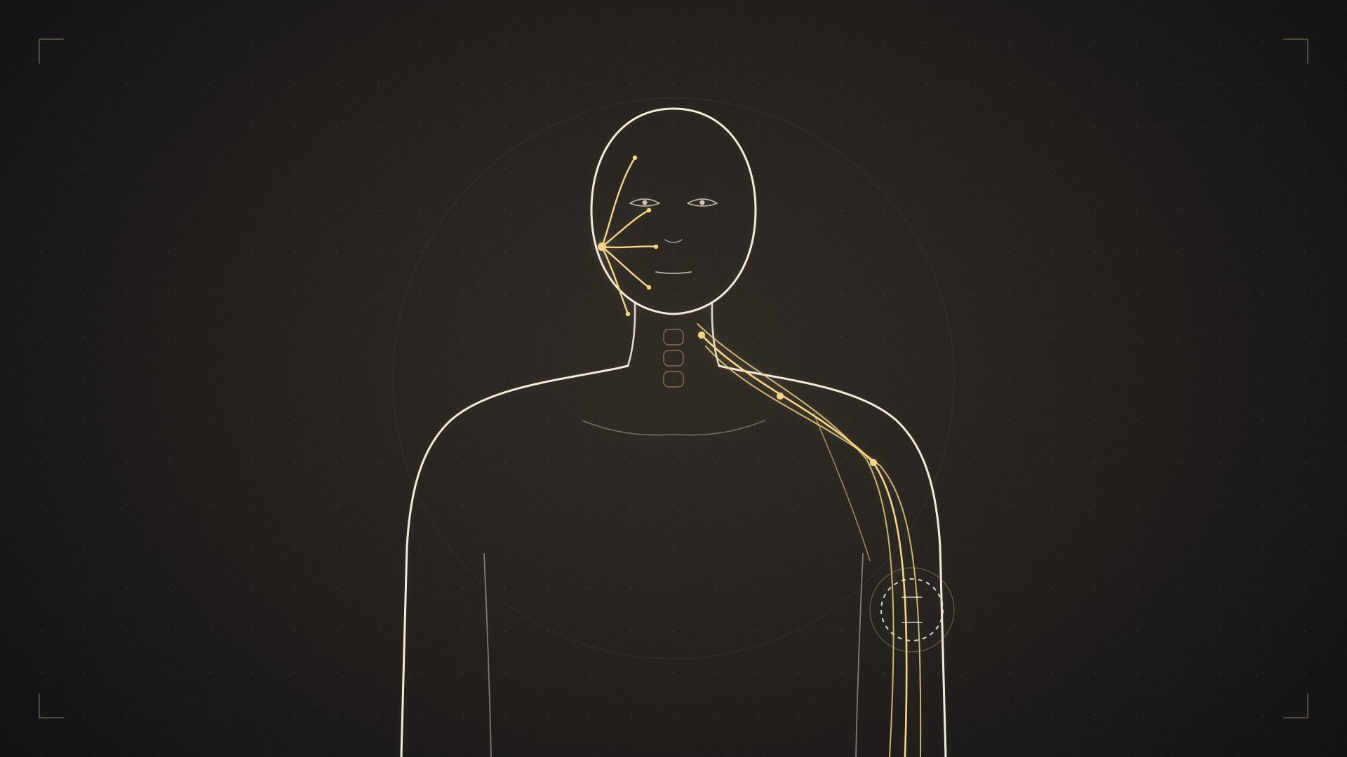
This article brings together in one place four major topics concerning the nerves of the face and arm. It can be read from start to finish, but most readers do not need to — if you have come with a specific problem, go straight to the relevant section using the table below.
| Section | Topic |
|---|---|
| Section 1 | Is it an emergency? Initial assessment |
| Section 2 | Why does time matter so much? (The backbone of this guide) |
| Section 3 | The facial nerve and facial paralysis |
| Section 4 | Facial paralysis that does not recover — reanimation surgery |
| Section 5 | Synkinesis — the problem of “too much or wrong movement” |
| Section 6 | The brachial plexus — the arm’s nerve network |
| Section 7 | Temporary weakness and facial paralysis after facelift surgery |
| Section 8 | Lessons common to all of them |
| Section 9 | Frequently asked questions |
Important note: This content is for general information and does not replace examination, diagnosis or treatment by a physician. If you have sudden weakness, loss of sensation or severe pain, seek medical care without delay.
Section 1: Is it an emergency? Initial assessment
1.1. If half of your face is not working: the forehead test
The most critical question in facial paralysis comes down to one sentence: is the problem in the brain, or along the path of the nerve?
The answer comes from a surprisingly simple anatomical fact: the forehead muscles are supplied by both halves of the brain. The lower half of the face, however, is supplied only by the opposite side.
| Peripheral paralysis (Bell’s, Ramsay Hunt) | Central paralysis (stroke) | |
|---|---|---|
| Wrinkling the forehead | Cannot — the forehead stays smooth | Can — the forehead wrinkles normally |
| Closing the eye | Does not close fully | Usually closes |
| Corner of the mouth | Drooping | Drooping |
| Arm/leg strength | Normal | Often weak |
| Speech | Pronunciation impaired but meaningful | Word-finding difficulty, meaningless speech |
Stand in front of a mirror and raise your eyebrows as if surprised.
-
If your forehead wrinkles on both sides and your mouth droops → it may be central: call 112
-
If the forehead on the paralysed side does not wrinkle at all → most likely a peripheral paralysis; not an emergency, but see a doctor the same day
1.2. Situations in facial paralysis that require calling 112 immediately
-
Weakness or numbness in an arm or leg
-
Speech disturbance, word-finding difficulty, not understanding
-
Sudden, severe headache
-
Loss of vision, double vision
-
Loss of balance, inability to walk
-
Difficulty swallowing, clouding of consciousness
-
Facial paralysis appearing after a head injury
-
Forehead muscles that keep working
In stroke treatment there is a narrow time window for clot-dissolving drugs. “I’ll see how it is in the morning” is an irreversible decision.
1.3. If your arm is not working: situations that require calling 112 immediately
-
Weakness in both arms at once — this is not a problem of the nerve network but of the spinal cord
-
Weakness in the legs as well, loss of bladder or bowel control
-
An absent pulse in the arm, a cold and pale hand — a vascular injury; hours matter
-
Rapidly growing swelling or bruising in the neck
-
The arm not working after a cut or stab wound
-
Arm paralysis together with shortness of breath
1.4. Not emergencies, but not to be delayed
-
Partial weakness or numbness of the arm after trauma
-
A newborn’s arm hanging limp at its side
-
Weakness following sudden severe shoulder pain
-
Facial or arm weakness that gradually increases over weeks — it does not look urgent, but it is the group that most needs investigation; tumours present this way
Section 2: Why does time matter so much? (The backbone of this guide)
If you read this section, everything else falls into place. Because underneath all these topics, from facial paralysis to the brachial plexus, lies the same biological fact.
2.1. Muscles cannot survive without nerves
A muscle that does not receive regular stimulation from its nerve begins to waste away. The points where the nerve connects to the muscle — the motor end plates — deteriorate irreversibly over time.
This deterioration is usually complete within 18–24 months. Beyond that point, even if you bring a perfect nerve to the muscle, the muscle will not respond, because the “socket” that would receive the signal no longer exists.
The entire logic of treatment follows from this single sentence:
-
If the muscle is still alive → bringing a nerve back to it is enough
-
If the muscle has died → a new muscle has to be brought in from elsewhere
2.2. How fast do nerves heal?
Torn nerve fibres can regrow inside an intact sheath. But their speed is fixed and slow: about 1 millimetre a day.
In everyday terms, this means:
-
From the neck to the biceps: about 8–10 months
-
From the neck to the hand: a year and a half or more
-
Distances are short in the facial nerve, which is why recovery in facial paralysis is usually faster
This is where the critical contradiction lies: while the nerve advances 1 mm a day, the muscle dies in 18–24 months. The nerve’s inability to reach a distant muscle in time explains why, in brachial plexus injuries, the hand is the hardest part to save.
2.3. The degree of damage and the chance of recovery
| Type of damage | What has happened | Recovery |
|---|---|---|
| Neurapraxia | The nerve is bruised or stretched, its structure intact | Recovers fully within days to weeks |
| Axonotmesis | The fibres are torn, the sheath intact | Slow, but sometimes possible |
| Neurotmesis | The nerve is completely severed | Does not heal on its own; surgery is needed |
| Avulsion | The root has been torn out of the spinal cord | Classic repair impossible; nerve transfer needed |
2.4. The general time window
The table below is the general framework that applies to both the face and the arm. Details are in the sections:
| Duration | Situation | Priority |
|---|---|---|
| 0–3 weeks | Diagnostic period | Too early for EMG; joint exercises and eye protection begin |
| 3 weeks–3 months | Mild damage recovers | EMG and imaging; the plan takes shape |
| 3–6 months | Decision period | If there is no recovery, surgery in this window |
| 6–12 months | The window is narrowing | Surgery is possible but results decline |
| 12–24 months | Critical threshold | Nerve surgery largely loses its value |
| After 24 months | No muscle | Muscle transfer, tendon transfer, static methods |
Two sentences follow from this:
One: if you are in the early period, “let’s wait a bit longer” is the most expensive advice. Getting an assessment is not agreeing to surgery — it simply keeps the door open.
Two: if years have passed, you are not too late. The path changes; the door does not close.
Section 3: The facial nerve and facial paralysis
3.1. What does the facial nerve do?
The seventh (the facial nerve) of the 12 pairs of cranial nerves leaving the brain controls the muscles of facial expression. But that is not its only job; it carries four separate functions in the same cable:
1) Moving the muscles of facial expression. Raising the eyebrows, closing the eyes, smiling, pursing the lips — about 20 muscles take their orders from it. The chewing muscles depend on another nerve; this is why in facial paralysis you struggle not with chewing but with keeping your mouth closed.
2) Regulating tear and saliva secretion. This is why the eye either waters excessively or dries out completely.
3) Taste from the front two-thirds of the tongue. The complaint “food tastes different, there is a metallic taste” is no coincidence.
4) Controlling the stapedius muscle in the inner ear. This muscle is a shock absorber that softens loud sounds. When it is out of action, sounds seem uncomfortably loud — hyperacusis.
3.2. Why does it get trapped so often?
The facial nerve passes through a very narrow bony canal (the Fallopian canal) inside the temporal bone, the hardest bone of the skull. Some parts of the canal fit the diameter of the nerve almost exactly.
The result: if the nerve swells for any reason, it has nowhere to go. The swollen nerve becomes trapped in its own canal, blood flow decreases and conduction stops. Think of it as the facial-nerve equivalent of carpal tunnel syndrome in the wrist.
This single fact explains why treatment starts with an anti-swelling drug (cortisone) and why acting early is critical.
3.3. Bell’s palsy — the most common cause (60–75%)
A sudden, one-sided peripheral facial paralysis for which no cause can be found is called Bell’s palsy.
“No cause can be found” is not quite accurate. Today the strongest explanation is this: the herpes simplex virus (HSV-1), which causes cold sores, lives silently for life in a nerve ganglion. It is reactivated by stress, fatigue, lowered immunity or an infection; the resulting inflammation makes the nerve swell and traps it in the bony canal.
Frequency: 20–30 cases per 100,000 people per year. The lifetime risk is about 1 in 60.
Who is affected more often: it is concentrated in the 15–45 age range; there is no difference between women and men or between right and left. Factors that increase the risk: diabetes (2–4 times), pregnancy — especially the last three months and the first week after delivery (about 3 times), pre-eclampsia, obesity, high blood pressure.
Classic course: it often starts with a dull pain behind the ear, then reaches its peak within hours, and at most within 72 hours. The defining feature of Bell’s palsy is that it is fast.
Warning sign: if facial paralysis progresses slowly over weeks, it is not Bell’s palsy. Slowly progressive paralysis suggests a tumour (parotid tumour, acoustic neuroma) and always requires imaging.
3.4. Ramsay Hunt syndrome (shingles of the ear)
It is the second most common cause, and it is critical not to confuse the two.
The virus from childhood chickenpox (VZV) lies dormant in nerve ganglia. If it reactivates years later in the ganglion of the facial nerve, Ramsay Hunt syndrome appears.
How it differs from Bell’s palsy:
-
Severe, burning pain — “like an electric shock”
-
Painful fluid-filled blisters on the outer ear, in the ear canal or inside the mouth — the most distinctive finding
-
Often accompanied by hearing loss, ringing in the ear and dizziness
-
Usually a more severe paralysis and a markedly worse prognosis
The blisters may appear before or after the paralysis, or not at all. If you see blisters in your ear, be sure to say so — antiviral treatment is unquestionably needed here and should be started early.
Prevention: over the age of 50, the shingles vaccine (recombinant form, 2 doses) markedly reduces the risk.
3.5. Other causes
Trauma: temporal bone fractures, facial injuries, birth trauma.
Ear diseases: chronic middle ear infection, cholesteatoma (a bone-eroding growth), severe outer ear infections. Facial paralysis with ear discharge means an ENT emergency.
Tumours: parotid tumours, acoustic neuroma, skull base tumours. Distinguishing features: slowly progressive paralysis, a lump in the face, recurrent paralysis on the same side.
Infections: Lyme disease (after a tick bite; an important cause of paralysis on both sides of the face in children), HIV, tuberculosis.
Diseases of the nervous system: Guillain-Barré, multiple sclerosis, sarcoidosis, Sjögren’s.
Paralysis on both sides: less than 1% of all cases and almost never simple Bell’s palsy. Lyme disease, Guillain-Barré, sarcoidosis, leukaemia/lymphoma, HIV and skull base fractures should be investigated. Because the face stays symmetrical, it is hard to notice; it stands out through an expressionless, mask-like face and inability to close the eyes.
3.6. Symptoms: not just a “drooping face”
Related to the facial muscles: drooping of the corner of the mouth, inability to wrinkle the forehead, inability to close the eye fully (lagophthalmos), the lower eyelid turning outwards, collapse of the side of the nose, biting the inside of the cheek, leaking drinks, difficulty with the sounds “P-B-M-F”, inability to whistle.
Not related to the facial muscles but dependent on the same nerve: pain behind the ear (often 1–2 days before the paralysis), hyperacusis, change in taste, dry eye or excessive watering.
A feeling of numbness in the face is common, but on examination there is no real loss of sensation — it is a deceptive feeling caused by the immobile muscle.
What should not be present (alarm if it is): hearing loss, severe dizziness, double vision, arm or leg weakness, fever, ear discharge, a lump in the neck, weight loss.
3.7. Diagnosis: which tests are needed, and which are not?
Bell’s palsy is a diagnosis of exclusion. The doctor assesses the speed of onset, the forehead muscles, the eardrum, the neck and parotid area, the other cranial nerves and the skin.
Severity of the paralysis — the House-Brackmann scale:
| Grade | Meaning |
|---|---|
| I | Normal |
| II | Mild — noticeable on close inspection; the eye closes fully |
| III | Moderate — obvious; the eye closes with effort |
| IV | Moderately severe; the eye does not close fully |
| V | Severe — barely perceptible movement |
| VI | Complete paralysis — no movement at all |
Roughly speaking, grade IV and above is a severe picture and requires closer follow-up.
The clear recommendations of international guidelines:
-
In typical Bell’s palsy, routine blood tests are not needed
-
In typical Bell’s palsy, routine MRI or CT is not needed — not having an MRI on the first day is not negligence but a guideline recommendation
-
Imaging is needed for: slowly progressive paralysis, no improvement at all after 3 months, recurrence on the same side, hearing loss or dizziness, trauma, ear discharge, a lump, paralysis on both sides
-
EMG: unnecessary in partial paralysis; in complete paralysis it can be done to estimate the chance of recovery
3.8. Treatment
Corticosteroids — the cornerstone of treatment. The single most important fact: they should be started within the first 72 hours. This is the recommendation with the highest level of evidence. They markedly increase the rate of full recovery; in about 1 in every 10 patients they turn the recovery from “partial” to “complete”.
Typical use is 50–60 mg of prednisolone a day, 5 days at the full dose and 5 days tapering. Your doctor sets the dose; do not stop on your own. If you have diabetes, be sure to say so — cortisone raises blood sugar and requires monitoring, but it does not rule out treatment. It can also be used during pregnancy.
If you have passed the 72 hours, the benefit decreases but is not zero; many doctors start treatment up to day 7.
Antiviral drugs. Clarity matters here: they should not be given on their own — this is an explicit “do not do” recommendation in the guidelines. They are considered together with cortisone, especially in severe and complete paralysis; there are data suggesting they reduce the risk of synkinesis. In Ramsay Hunt syndrome, however, antivirals are always given; there is no debate.
Eye care — the most neglected and most important issue. The most lasting damage from Bell’s palsy occurs not in the face but in the eye.
If the eyelid does not close fully, the cornea is constantly exposed; it dries, gets scratched and becomes infected. What is more, two protections are lost at once: the lid does not close and tears decrease.
-
By day: preservative-free artificial tears, every hour or as often as needed. Glasses outdoors
-
At night: eye gel/ointment + gently closing the lid with hypoallergenic tape (the tape goes on the lid, not on the lashes)
-
See an eye doctor immediately for: redness, pain, a gritty feeling, blurred vision, sensitivity to light
Physiotherapy. The muscles are intact; the problem is in the nerve; exercise does not speed up the nerve. In mild cases the added benefit is unclear. In severe paralysis (V–VI) or paralysis lasting more than 3 months it is recommended — the aim is to prevent muscle shortening and limit faulty nerve repair. The most effective approach is slow, controlled, isolated movements in front of a mirror; rough straining may increase the risk of synkinesis.
Unproven or risky: electrical stimulation (there are data on potential harm), acupuncture, hyperbaric oxygen, low-level laser, vitamin B. These should not replace cortisone and eye care or cause delay.
Surgical decompression. Highly controversial; it is considered in selected patients with complete paralysis and severe loss on nerve conduction tests, within the first 2 weeks, in experienced centres. It is not a routine treatment.
3.9. Recovery timeline
First 72 hours: the paralysis may get worse — this is normal and does not mean the treatment is not working.
Weeks 1–3: the first flickers of movement in most patients. Usually eye closure and movement of the corner of the mouth return first.
Weeks 3–8: about 70–85% of patients recover almost completely during this period.
Months 3–6: gradual improvement in severe cases; signs of synkinesis may appear in this period.
Months 6–12: recovery slows. The picture after month 12 is generally considered permanent.
Good prognosis: partial paralysis, early treatment, young age, recovery that begins quickly, no pain. In children and pregnant women, full recovery is close to 90%.
Difficult prognosis: complete paralysis, older age, diabetes, high blood pressure, severe pain, Ramsay Hunt syndrome, no movement at all in the first 3 weeks.
Recurrence: about 6–7%; the average interval between two episodes is 10 years. Recurrent paralysis on the same side always requires imaging.
Read more: A detailed guide to facial paralysis
Section 4: Facial paralysis that does not recover — reanimation surgery
4.1. Is there a “definitive cure”? An honest answer
One of the most searched phrases on the internet is “a definitive cure for facial paralysis”. Let us answer clearly:
There is no “definitive cure” that returns the paralysed nerve to its former state. Bringing a dead nerve back to life or restoring a wasted muscle is not possible with today’s medicine. Do not trust approaches that promise this.
But permanent facial paralysis has permanent solutions. Facial reanimation surgery can restore the smile by bringing a new nerve supply or a new muscle to the paralysed side; it can protect the eye, provide resting symmetry and improve eating and speaking.
The goal is not “getting your old face back” but “gaining a face that works.”
4.2. Why does facial paralysis become permanent?
After tumour surgery — one of the most common causes. In operations for acoustic neuroma, skull base tumours and parotid cancer, the nerve may not be preservable or may be deliberately sacrificed. This group is the most favourable for reanimation: the site and time of the cut are known, and repair can be planned in the same session.
Severe Ramsay Hunt syndrome. Trauma. Cholesteatoma and chronic ear disease. Congenital conditions (Möbius syndrome). Bell’s palsy that was treated late or ran a severe course. Systemic diseases (sarcoidosis, advanced Lyme disease).
4.3. The treatment map
| Your situation | Leading options |
|---|---|
| The nerve was cut during surgery, the ends are available | Direct repair or graft — in the same session, best result |
| Paralysis less than 12 months old, muscles alive on EMG | Masseter transfer, hypoglossal transfer, cross-facial graft |
| Paralysis more than 2 years old, muscles wasted | Gracilis muscle transfer (smile surgery) |
| Does not want or cannot tolerate surgery | Static sling, brow lift, eye procedures |
| The eye does not close | Upper lid weight, lower lid tightening |
| Congenital paralysis | Muscle transfer; in two stages if both sides |
| Child patient | Wider window; nerve surgery up to 18 months |
These options are not rivals. In most patients treatment is staged: the eye is protected first, then a muscle transfer is done for the smile, and finally a static procedure or Botox is added for symmetry.
4.4. Nerve operations
Direct repair and grafting. If the nerve was cut recently and its ends can be found, they are joined; if there is a gap, a sural nerve taken from the leg is placed as a bridge. The best results come from repair in the same operation or in the first months.
Masseter (chewing nerve) transfer. One of the most widely used methods today. A branch of the nerve that drives the chewing muscle is connected directly to the smile branch of the facial nerve.
Why it is preferred: it is fast (first movement at about 5 months on average), it is strong (it carries more than ten times as many fibres as the alternatives — about 1,500 versus 100–200), no graft is needed in between, and there is little loss at the donor site.
Disadvantage: the smile is not spontaneous at first; the patient first learns to smile by “clenching the teeth”. It becomes more natural over time with rehabilitation, but in some patients this may persist.
Hypoglossal (tongue nerve) transfer. Part of the nerve that moves the tongue is redirected to the facial nerve. Because modern techniques use only part of the nerve, the tongue wasting and speech and swallowing problems of the past have largely disappeared.
Accessory (shoulder nerve) transfer. Part of the nerve to the trapezius muscle, which is responsible for shrugging the shoulder, is taken. It is usually used with muscle transfer. In my own practice I use it frequently because of its advantage of producing few involuntary contractions.
Cross-facial nerve grafting (CFNG). One of the surplus branches of the facial nerve on the healthy side is carried to the paralysed side through a long graft passed beneath the upper lip. In this way both sides are fed by the same command.
Advantage: it is the only method that can provide a genuinely spontaneous smile.
Disadvantages: it is slow (6–8 months to cross the 20 cm graft), lossy (only 20–50% of fibres cross at each connection), can be unpredictable when used alone, and success falls with age.
Combination (the “babysitter” technique). The modern trend is to combine the two: the masseter transfer quickly keeps the muscles alive, while fibres from the cross-facial graft slowly arrive and over time take over spontaneous movement.
4.5. Muscle transfer — “smile surgery”
If the muscles can no longer be revived, the solution is to bring in a new muscle from elsewhere.
How it is done: part of the gracilis muscle on the inner thigh is taken with its own vessels and nerve and moved to the face; its vessels are connected to facial vessels and its nerve to the chosen donor nerve using microsurgery. One end is fixed to the corner of the mouth, the other to the cheekbone. The operation usually takes 6–8 hours.
Which nerve will drive the muscle?
| Donor nerve | Strength | Weakness |
|---|---|---|
| Masseter nerve | Strong, reliable, fast | Voluntary at first — “smiling by biting” |
| Accessory nerve | Strong, reliable, fast, fewer involuntary movements | Voluntary at first — “smiling by lifting the shoulder” |
| Cross-facial graft | Spontaneous, natural smile | Weaker and slower, may be unpredictable |
| Dual innervation | Combines the advantages of both | Complex, longer operation; the masseter input may dominate |
In recent years, dual innervation and multi-vector designs (directing the muscle towards the corner of the mouth, the upper lip and the lower eyelid) have become widespread.
The process: surgery → no movement for the first 3 months → first contractions in months 3–9 → increasing strength and rehabilitation in months 9–18 → final result in months 18–24.
How much movement can be expected? The result is measured by how many millimetres the corner of the mouth moves. Around 1 cm is a good result; under 4 mm is considered inadequate. Adjusting the muscle’s length may require additional operations.
4.6. Static methods
They do not restore movement, but they provide resting symmetry and correct functional problems.
Fascial sling: the corner of the mouth is suspended upwards with a strip of connective tissue taken from the thigh; leaking and drooling decrease. The effect is immediate.
Brow lift: prevents a drooping eyebrow from obstructing the field of vision.
One-sided facelift: repositioning of sagging cheek tissue.
Nasal ala suspension: a collapsed side of the nose makes breathing difficult; suspension opens the nasal passage — a gain patients do not expect but are very pleased with.
4.7. Eye surgery — always a priority
Upper lid weight (gold implant). The most commonly used method. A thin plate is placed inside the lid, and gravity closes it. Simple, reversible, effective. Possible problems: the implant being visible, shifting, rarely extruding, mild astigmatism.
Lower lid tightening (lateral tarsal strip). The loose, outward-turned lower lid is tightened and seated against the eyeball.
Tarsorrhaphy. Stitching part of the lids together; usually for temporary, emergency protection.
And in every case: drops by day, gel and tape at night, glasses outdoors.
Read more: Treatment options for permanent facial paralysis
Section 5: Synkinesis — the problem of “too much or wrong movement”
Permanent facial paralysis also has an opposite: the nerve has healed, but healed wrongly.
5.1. Why does it happen?
As the nerve heals, its fibres grow again and connect to the muscles — but not always to the right address. A fibre meant for an eye muscle may go to a mouth muscle, and one meant for a mouth muscle may go to the eye. Like a wrongly soldered circuit.
About one in four people who have had facial paralysis show some degree of synkinesis a year later.
5.2. What does it look like?
-
Oral-ocular synkinesis: the eye narrows or closes when smiling
-
Ocular-oral synkinesis: the corner of the mouth twitches when blinking
-
Crocodile tears (Bogorad syndrome): the eye waters when eating — fibres meant for the salivary gland have connected to the tear gland
-
Contracture: the paralysed side stays tight; at rest it may look even more “pulled” than the healthy side — it can become unclear which side is paralysed
Synkinesis usually begins in months 3–6, becomes more obvious over time and does not go away completely on its own.
5.3. Treatment
The aim is not to add movement but to silence excess and wrong movement.
Botox. The mainstay of treatment for this group. It is injected in small doses into overactive muscles; the effect lasts 3–6 months and is repeated. It is sometimes also applied to the healthy side to achieve balance.
Neuromuscular retraining. A special physiotherapy programme, using a mirror and biofeedback, that teaches you to suppress wrong contractions and isolate the correct movement. It gives the best result combined with Botox — Botox turns down the noise, and rehabilitation teaches the correct movement.
Selective neurectomy and myectomy. In resistant cases, the nerve branches going to the overactive muscle are cut selectively, or part of the muscle is removed. These operations reduce, but do not completely eliminate, the need for Botox.
Section 6: The brachial plexus — the arm’s nerve network
6.1. Your arm is actually controlled from your neck
All the nerves that move your arm and give it sensation come not from the arm itself but from the cervical spine. Five nerve roots leaving the neck form a complex network beneath the collarbone and give rise to the major nerves of the arm. This network is called the brachial plexus.
Levels: roots (C5, C6, C7, C8, T1) → trunks (upper, middle, lower) → divisions and cords → terminal nerves.
| Nerve | What it does | If it fails |
|---|---|---|
| Axillary | Lifts the shoulder sideways | The arm cannot be raised sideways |
| Musculocutaneous | Bends the elbow | The elbow cannot bend |
| Radial | Lifts the wrist and fingers | “Wrist drop” |
| Median | Thumb and grasping | Grasp is impaired |
| Ulnar | Opening and closing the fingers, fine movement | “Claw hand”, hand muscles waste |
Why is it fragile? The nerves are like a taut bridge between the neck and the arm; if the head is forced one way and the shoulder the other, they stretch like elastic and tear. They also pass through the narrow passage between the collarbone and the first rib.
A critical anatomical detail: the upper roots (C5–C6) are anchored to the spine by strong ligaments, so they usually rupture outside the spine. The lower roots (C8–T1) lack this support and can be torn directly out of the spinal cord — avulsion.
6.2. Preganglionic or postganglionic?
This is the distinction surgeons care about most:
-
Postganglionic: the rupture is outside the spine. It can be repaired and a graft placed. Better prognosis.
-
Preganglionic (avulsion): the root has separated from the spinal cord. There is nowhere to attach a graft. Poor prognosis; nerve transfer is needed.
Findings that suggest avulsion: Horner syndrome (drooping eyelid, smaller pupil and loss of sweating on that half of the face, on the same side), winging of the shoulder blade, severe burning, electric-like pain inside the arm, shortness of breath, weakness in the neck muscles. These can be hard to interpret as a patient. A detailed examination and further tests are needed.
6.3. The root map and classic patterns
| Root | Movement lost | Numb area |
|---|---|---|
| C5 | Raising the shoulder sideways, external rotation | Over the shoulder, outer side of the arm |
| C6 | Bending the elbow, lifting the wrist | Outer forearm, thumb |
| C7 | Straightening the elbow, lifting the fingers | Middle finger |
| C8 | Bending the fingers, grasping | Ring and little fingers |
| T1 | Small muscles of the hand | Inner forearm |
-
Upper type (C5–C6, “Erb’s”): the shoulder and elbow do not work, the hand works. The “waiter’s tip” position. The most common type, with the best prognosis.
-
Lower type (C8–T1, “Klumpke’s”): the shoulder and elbow work, the hand is paralysed. Claw hand. Rare on its own.
-
Total type (C5–T1): the whole arm is paralysed. The most severe picture.
Pain deserves special mention. In avulsion, there can be unbearable pain in an arm you cannot feel — the brain generates its own pain for a region from which it receives no signals. It does not respond to ordinary painkillers; neuropathic pain medication is needed. Patients often think this pain is “imaginary” and do not mention it.
6.4. Causes
Birth-related injury. 0.2–5 cases per 1,000 live births. The greatest risk factor is shoulder dystocia, followed by a large baby (over 4 kg) and breech delivery. Note: in large babies the risk persists even with caesarean delivery.
Distribution by type: 60% upper type, 20–30% extended upper type, 10–20% total.
The most important information: about 60% of babies recover without surgery, and most of the recovery happens in the first 3–4 months.
The surgical decision: if the hand does not work at all and there is no improvement by months 2–3, early surgery; if elbow bending has not returned by months 3–6, nerve surgery; if the “cookie test” (bringing the hand to the mouth) fails at 9 months, surgery. Nerve surgery is usually performed in the 3–6 month period; muscle and tendon operations after the age of 2–3.
For families: even if surgery is not performed, physiotherapy is essential from the first days — it does not speed up nerve recovery, but it prevents the joints from freezing. The most common lasting problems: internal rotation contracture of the shoulder (close to 70%), forearm and elbow contractures, glenohumeral dysplasia, and a shorter arm. Follow-up extends over years.
Trauma in adults. The most common scenario is a motorcycle accident. This group accounts for the most severe pictures; more than one root is often affected and the rate of avulsion is high.
The timing rule: the first 3–6 months are ideal for surgical exploration and repair; results decline at 6–9 months; after 9–12 months, nerve repair is generally not recommended — the options shift to tendon transfer and free muscle transfer. In a cut injury, if the nerve end is identifiable, it is repaired immediately, without waiting.
Parsonage-Turner syndrome (brachial neuritis). There is no trauma, but the plexus is affected. 1–3 cases per 100,000 people per year.
The classic course has two phases: (1) sudden, very severe shoulder and arm pain — it often starts at night and lasts days to weeks. (2) As the pain eases, weakness appears; the shoulder blade may wing and muscle wasting is seen.
Triggers: viral infection, recent surgery, vaccinations, pregnancy, heavy exercise. In most cases no clear cause is found; there is also a hereditary form.
Why is it confused with other conditions? It is mistaken for “frozen shoulder”, a rotator cuff tear or a herniated cervical disc. The distinguishing clue: the pain comes first and the weakness later, and the weakness is disproportionately marked compared with the severity of the pain.
Treatment: pain control in the early period; corticosteroids started in the first 1–2 weeks may shorten the duration of pain; then physiotherapy. The prognosis is generally good but slow — about a third of patients show marked improvement in the first year, and the great majority within three years.
Thoracic outlet syndrome (TOS). Not a rupture but compression. Numbness that increases when the arm is raised (especially in the little and ring fingers), rapid fatigue of the hand, neck and shoulder pain. Risk factors: a cervical rib, poor posture, repetitive overhead work, a heavy backpack. Treatment is almost always physiotherapy first; surgery is considered in patients who do not respond and have objective nerve findings. The diagnosis is controversial; the decision to operate should not be rushed.
Sports injury (“stinger”). Common in contact sports. A burning electric sensation in the arm lasting seconds to minutes and temporary weakness; usually neurapraxia, which resolves completely. However: if the symptoms are in both arms, it is not a stinger — it may be a spinal cord injury and is an emergency. Recurrent stingers suggest narrowing of the cervical canal.
Tumours and radiation. A Pancoast tumour, arising at the apex of the lung, can press on the lower roots and cause slowly progressive arm pain, weakness of the hand and Horner syndrome. Radiation plexopathy, on the other hand, can appear months to years after treatment and, unlike tumour involvement, is usually less painful.
6.5. Diagnosis
EMG should be done 3–4 weeks after the injury — if done earlier, it misleads. An EMG at 3 months gives a clearer result and is repeated at 3 months; early signs of regeneration on EMG appear weeks before visible movement and change the surgical decision.
MRI shows the plexus, swelling, masses and the pseudocysts that signal avulsion. CT myelography is among the most reliable methods for showing root avulsion. Ultrasound shows nerve constrictions, especially in Parsonage-Turner syndrome. Plain X-rays show fractures, a cervical rib and an elevated diaphragm.
6.6. Treatment
From day one, for everyone: range-of-motion exercises (even if the nerve recovers, a frozen shoulder is of no use), an arm sling and correct positioning, protection of the insensate hand (burns and cuts go unnoticed; daily visual checks are essential), swelling and skin care.
Pain management: nerve pain medications such as gabapentin and pregabalin, some antidepressants, nerve blocks; special surgical procedures for resistant avulsion pain. Psychological support is part of treatment.
Surgery:
-
Nerve grafting — if the rupture is outside the spine; a short graft (under 10 cm) markedly improves results
-
Nerve transfer — in avulsion. The Oberlin transfer (a small fascicle from the ulnar nerve to the biceps nerve) gives gratifying results for elbow bending; accessory nerve to suprascapular nerve for the shoulder; intercostal transfers
-
Free muscle transfer — the gracilis muscle from the leg in late or very severe cases
-
Tendon transfers and arthrodesis — in cases too late for nerve repair
Rehabilitation: surgery is a beginning. While the nerve regrows, physiotherapy keeps the joints ready; when the muscle starts to contract, the brain has to learn the new connection. For example, after an Oberlin transfer the patient first bends the elbow with a “squeeze your hand” movement; over time the brain separates the two.
An honest sentence: in severe and total injuries, the goal is usually not “the arm being as it was before” but gaining shoulder stability, elbow bending and, if possible, simple grasping. Regaining the fine functions of the hand is the area where today’s surgery struggles most.
Read more: Brachial plexus injury guide
Section 7: Temporary weakness and facial paralysis after facelift surgery
At first glance this section may seem disconnected from the others. It is not: the technical difficulty of facelift surgery stems precisely from the need to protect the facial nerve. It is this nerve that determines the layer in which the surgeon works and where their attention goes.
7.1. The relationship between facelift surgery and the facial nerves
The facial nerve controls most of the face’s expressive functions, from forehead movements and eye closure to smiling and the movement of the lower lip and neck muscles. Because the techniques used in facelift surgery proceed in different anatomical layers, their relationship with the branches of the facial nerve also differs.
7.2. Does the facelift technique affect the risk of nerve injury?
Deep plane facelift is one of the facelift methods that must be performed with the greatest technical care, because it works in an anatomical layer closer to the branches of the facial nerve. Especially in the areas where the face’s retaining ligaments are released, the surgeon may come close to the nerve branches; in some operations these branches can be seen and directly protected.
At first glance this may give the impression that deep plane facelift is riskier. However, working close to the nerves does not necessarily mean less controlled surgery for an experienced surgeon. A surgeon who masters the anatomical layers can proceed knowing which tissue they are releasing and where the nerves may lie. Seeing the nerve can, in some cases, even make it easier to protect it in a controlled way.
Conversely, methods considered more superficial, such as SMAS plication, are not entirely risk-free either. Sutures placed out of view and deeper than necessary can compress nerve branches or catch them in the stitch. So the name of the technique alone is not enough to determine nerve safety.
Current studies do not show a clear, definite difference in facial nerve injury between deep plane and other SMAS techniques. That said, direct comparative studies are limited. This is why neither absolute statement — “deep plane is definitely more dangerous” or “it is definitely safer because the nerve is seen” — is correct. The real determinants are the surgeon’s mastery of facial anatomy, their experience with the technique they use and careful dissection.
7.3. How often does permanent facial paralysis occur after a facelift?
Permanent motor nerve damage after a facelift is extremely rare. Recent large reviews report permanent motor nerve injury at around 0.05%, that is, less than one in a thousand. In deep plane facelift studies, temporary motor nerve involvement is around 1–3%, while permanent injury is much rarer.
That said, not every asymmetry seen in the early period after surgery means nerve injury. Swelling, the effect of local anaesthesia, uneven swelling of tissues, work on the platysma and neck muscles, or temporary stretching of the nerve can create asymmetry, especially of the lower lip. After extensive and combined operations, such early asymmetries are more common than true nerve injury, and the great majority resolve within weeks or months.
This is why temporary weakness, early asymmetry and permanent nerve damage must be assessed separately.
7.4. In which patients are temporary weakness and asymmetry more likely?
The likelihood of temporary weakness or marked asymmetry may increase in the following situations:
-
A previous face or neck lift
-
Second or third facelift operations
-
Adhesions in the tissues caused by previous thread lifts, Endolift, radiofrequency or similar energy treatments
-
Heavy filler treatments that may affect the surgical layers
-
Recent Botox preventing full assessment of muscle movements before surgery
-
The need for extensive platysma and deep neck surgery because of advanced neck sagging or a large, heavy neck
-
Adding procedures to the facelift that may increase swelling, such as fat grafting, laser or similar
-
Long and multiple surgical procedures
-
The surgeon not sufficiently mastering the technique used and the anatomy of the facial nerve
Some of these factors increase the risk of true nerve injury, while others only accentuate postoperative swelling and temporary asymmetry. Temporary weakness can be seen even after an operation by an experienced surgeon; however, the risk of permanent damage is very low.
7.5. What can be done if the nerve is found to have been cut during surgery?
If a cut in a branch of the facial nerve is noticed, the most appropriate approach is to repair it during the same operation if possible. The nerve ends are brought together under a microscope or high magnification, with very fine microsurgical sutures and without tension. If the ends cannot be joined directly, a nerve graft may need to be placed in between.
It is important that this repair is carried out by a surgeon experienced in peripheral nerve surgery and microsurgery. Correct repair at the moment the injury occurs gives the best chance of recovery. Even so, nerve healing is slow, and full symmetry, completely natural movement or recovery without synkinesis cannot be guaranteed.
7.6. When should you worry about weakness after a facelift?
Many mild, limited asymmetries can be monitored in the early period. However, an earlier assessment by a surgeon experienced in facial nerve surgery is needed in the following situations:
-
Complete or severe loss of movement appearing immediately after surgery
-
Suspicion that the nerve may have been cut or seriously injured during surgery
-
Surgery that was technically difficult or problematic
-
A second or third facelift
-
Surgery in a heavily scarred field with disrupted anatomical layers
-
Previous thread lifts, Endolift, radiofrequency or other energy treatments
-
The eye not closing fully
-
Weakness that is getting progressively worse
-
No sign of improvement at all within weeks
-
No marked improvement by the third month
If there is serious suspicion that the nerve was cut, waiting three months is not right. In this case, the operating surgeon should be consulted and an early facial nerve surgery assessment carried out.
Needle EMG can begin to show whether there is degeneration in the nerve about 2–3 weeks after the injury. In the following weeks it helps assess signs of reinnervation. However, it is not necessary for every mild weakness and asymmetry. If there is no marked clinical improvement at three months, the examination and EMG findings should be assessed together to form the next treatment plan.
7.7. Why is facial paralysis after a facelift difficult to assess?
There is an important difficulty that sets facial paralysis after a facelift apart from other causes: the person who best knows where and by what mechanism the nerve may have been affected is the surgeon who performed the operation.
A temporary nerve involvement after an operation in which every step went correctly in the hands of an experienced surgeon, and a serious nerve injury occurring in a difficult operation in which the anatomical layers were lost, can look similar from the outside in the early period. It is not always possible to tell with certainty from a single examination whether the nerve was merely stretched, thermally affected, compressed under a suture or completely cut.
This is why the following information should be considered together in the assessment:
-
Whether the weakness appeared immediately after surgery or later
-
Which facial movements are affected
-
Whether the nerve was seen during surgery or any problem occurred
-
The operative report and, if possible, intraoperative images
-
Serial examinations, standard photographs and movement videos
-
EMG performed at the right time
-
In selected patients, high-resolution ultrasound or MRI aimed at nerve imaging
-
Direct communication with the surgeon who performed the operation
Not every asymmetry after a facelift is permanent facial paralysis. But simply waiting is not right either for patients with a suspected serious nerve injury. Distinguishing in time between temporary involvement and an injury that may need repair is the most important step of treatment.
In experienced hands, facelift operations are very safe surgery as far as the nerves are concerned. For more detailed information on the deep plane facelift technique, appropriate patient selection, the other risks of surgery and the recovery process, see the Deep Plane Facelift Guide.
Read more: Deep plane facelift guide
Section 8: Lessons common to all of them
Although the four topics look different, they come down to the same five principles.
1. Time comes before technique. 72 hours in facial paralysis, 3–6 months in the brachial plexus, 12–24 months in reanimation. In all of them, “let’s wait a bit longer” is the most expensive sentence. Getting an assessment is not agreeing to surgery.
2. Function comes before aesthetics. In facial paralysis, the eye always comes first; the most lasting damage happens there and it is completely preventable. In the brachial plexus, protecting the insensate hand has the same priority.
3. Rehabilitation is half the operation. When the nerve is reconnected, the brain has to learn the new connection. This does not happen on its own.
4. Any slowly progressive paralysis is suspicious. In the face and in the arm alike: weakness increasing over weeks to months suggests a tumour and requires imaging.
5. The surgeon comes before the technique. All these fields are practised in a small number of centres. The questions to ask are the same: What is your experience with this operation? Can I see the results of your own patients? How many stages will the process involve? Who will run the rehabilitation? How will things work if there is a complication?
Warning signs — common to all: approaches that guarantee the result, promise a “definitive cure”, gloss over the risks, reduce the process to a single operation or never mention rehabilitation.
Section 9: Frequently asked questions
Is facial paralysis a stroke?
Mostly not. If the forehead wrinkles and the mouth droops, it may be central — call 112. If the forehead does not wrinkle, it is most likely a peripheral paralysis.
How many days does facial paralysis take to go away?
About 70–85% of patients recover almost completely within 3–8 weeks. In severe cases the process can extend to 6–12 months.
I started cortisone late — will it still help?
After 72 hours the benefit decreases but is not zero; many doctors start treatment up to day 7.
Did it happen because I was exposed to a draught?
No. Sleeping in front of an air conditioner or an open window does not cause Bell’s palsy — it is a culturally widespread belief with no scientific basis. (Still, direct wind on an eye that does not close is genuinely bad for the cornea.)
Will my face heal faster the more I work it?
No. The nerve heals at its own pace; however, physiotherapy and exercises are very important for better results in moderate to severe paralysis.
I have had paralysis for 5 years — am I too late?
Probably yes for nerve surgery, but not for muscle transfer. A smile can be restored even years later.
My arm has no feeling at all but hurts a lot — is that possible?
Yes, and it is typical. The part of the brain that receives no signals generates its own pain; neuropathic pain treatment is needed.
My MRI was clear — does that mean nothing is wrong?
No. A normal MRI does not completely rule out nerve damage. Examination and further tests such as EMG are also important.
When should I worry about my baby’s arm?
About 60% recover on their own, but for those who do not, the decision window is 3–6 months. Do not neglect follow-up.
Is a birth injury the doctor’s fault?
Not always. Shoulder dystocia is an unpredictable emergency; in large babies it can occur even with caesarean delivery.
Does smoking affect recovery?
Yes, in all of these conditions. It directly threatens the success of nerve repair and grafts, and vessel patency in microsurgery.
Do stem cells or nerve-regenerating treatments work?
Research is ongoing, but today there is not enough evidence to support their routine use. Postponing proven treatments in search of experimental options costs time — and in these conditions, time translates directly into results.
Are nerve problems after a facelift temporary?
Usually, but not always. Your surgeon will give the most accurate answer. If the right plan was followed during surgery and the operation went smoothly, the likelihood that it is temporary is high. If you cannot get clear answers, it is advisable to get an opinion from a doctor who works in nerve surgery.
Are these operations covered by insurance?
Reanimation after paralysis and nerve surgery are reconstructive procedures; coverage varies by insurer and indication.
Summary: 10 things to remember
-
The forehead test. If your forehead wrinkles and your mouth droops — call 112. If it does not wrinkle, the picture is most likely benign.
-
The 72-hour rule in facial paralysis. The window for cortisone is narrow and does not come back.
-
The eye always comes first. The most lasting damage happens there, and it is completely preventable.
-
Blisters in the ear may mean Ramsay Hunt syndrome. An antiviral is urgently needed; do not forget to mention it.
-
Without a nerve, muscles waste irreversibly in 18–24 months. All the timing rules follow from this.
-
In the brachial plexus, the decision window is 3–6 months; after 9–12 months, nerve repair loses its value.
-
Do not have the EMG done before 3–4 weeks — if done early, it misleads.
-
Even if you are late, the door does not close. Muscle transfer and static methods can be used even years later.
-
Slowly progressive paralysis is suspicious — in the face and in the arm alike; it requires imaging.
-
The surgeon comes before the technique. Experience, patient selection and rehabilitation matter more than the name of the method.
For a personal assessment of your situation, you can request an evaluation appointment; for the physician’s training and experience, see the physician profile.
This content is for general information and does not replace examination, diagnosis or treatment by a physician. If you notice sudden weakness, loss of sensation or severe pain in your face or arm, seek medical care without delay.
Assoc. Prof. Ahmet Hamdi Sakarya, MD, MSc
Specialist in Plastic, Reconstructive and Aesthetic Surgery
Assoc. Prof. Ahmet Hamdi Sakarya completed a two-year fellowship in reconstructive microsurgery and peripheral nerve surgery at Chang Gung Memorial Hospital in Taiwan, one of the world’s leading high-volume microsurgery centers. His clinical practice focuses on facial paralysis and facial reanimation, brachial plexus and other peripheral nerve injuries, functional muscle transfers, free tissue transfers and facial aesthetic surgery.


