

Tissue that brings its own blood supply: types of flaps, how they differ from grafts, and which one is chosen in which situation
Introduction: Beyond simply closing a wound
Some wounds cannot be closed with stitches. Some cannot be closed even with a skin graft. If bone is exposed, a tendon is visible, a hip prosthesis has become uncovered, or tissue that has received radiotherapy has failed to heal for months, there is only one solution: bringing living tissue to the area that carries its own blood supply with it.
This tissue is called a flap, and it forms the backbone of reconstructive surgery.
Flap surgery is the broadest and most technical field within plastic and reconstructive surgery. It covers a spectrum ranging from closing a simple incision on the cheek to rebuilding the jawbone with bone taken from the leg.
This guide explains flap surgery in language a patient can understand: what a flap is, how many types there are, how it differs from a graft, which one is chosen in which situation, and how recovery works.
Important note: This content is for general information only and does not replace examination, diagnosis or treatment by a physician.
1. What is a flap?
Definition: A piece of tissue that carries its own blood supply (the artery and vein that nourish it) along with it.
This is the key concept: its own vessels. When a flap arrives at its new location, it does not depend on the underlying tissue for nourishment; it brings its own blood.
What can it contain? Skin alone; skin and fat; skin, fat and muscle; muscle alone; bone; or a combination of these. A nerve can be included when needed — in that case the flap can have sensation or movement.
The difference between a graft and a flap
This distinction is the key to understanding reconstructive surgery.
| Graft | Flap | |
|---|---|---|
| Own blood supply | No | Yes |
| How it survives | Nourished by the underlying bed | Through its own vessels |
| Requirement for the bed | A well-vascularized bed is essential | Can be placed on a poor bed |
| Thickness | Must be thin | Can be thick and bulky |
| Over exposed bone/tendon | Cannot be used | Can be used |
| On irradiated tissue | Usually does not take | Can be used |
| Technical difficulty | Low to moderate | Moderate to very high |
The decision rule is simple: If the wound bed has a good blood supply and thin coverage is enough, a graft; if the bed is poor, bone/tendon/an implant is exposed, or volume is needed, a flap.
2. Types of flaps: Four different classifications
Flaps are classified from four different perspectives, and these classifications overlap. A single flap can be “local, axial and fasciocutaneous” at the same time.
2.1. By distance
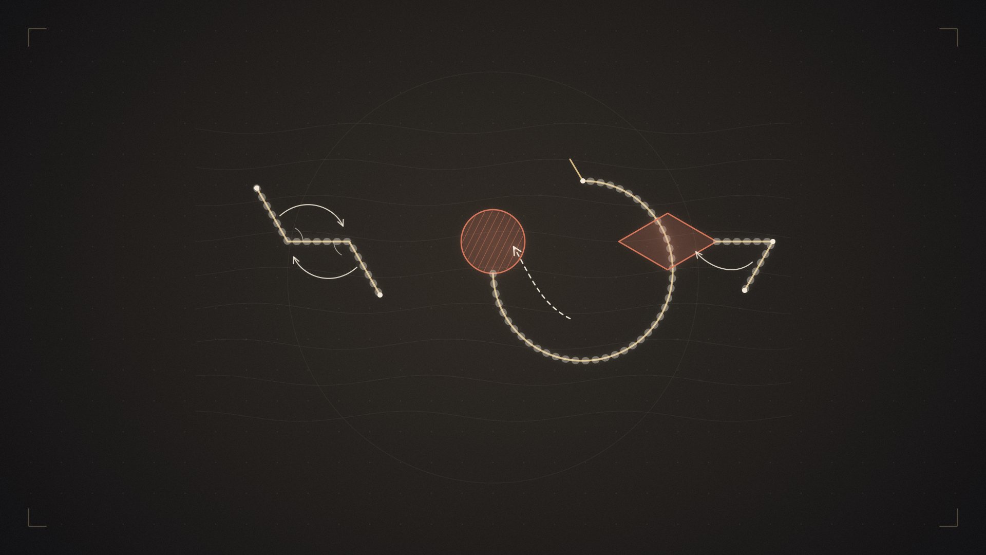
Local flap. Prepared from tissue immediately next to the wound and moved into the defect. The skin around the incision is advanced, rotated or swapped. It is the most commonly used type of flap in facial surgery — because the surrounding skin provides the best match in color and texture.
Examples: advancement flap, rotation flap, transposition flap, Z-plasty, rhomboid flap, bilobed flap.
Regional flap. Taken from the general region of the wound, but from a little farther away. It is moved by rotating it on a specific vessel.
Examples: a flap moved from the forehead to the nose (the paramedian forehead flap — the classic for nasal reconstruction), a flap moved from the chest muscle to the neck, a flap moved from the calf muscle to the knee.
Distant flap (pedicled). Tissue is taken from a distant area and “carried” to the wound while remaining attached to its vessel. In hand injuries, the hand is stitched to the abdomen so that it is nourished there, and it is separated after 2-3 weeks. It is used less today because of free flaps, but it is still valuable in some situations.
Free flap. Its vessel is divided, the tissue is moved to a distant area and sewn to new vessels under the microscope. There is no distance limit. This is a broad topic in its own right and is covered in a separate guide.
2.2. By blood supply
Random pattern flap. It has no specific named vessel; it is nourished by the capillary network beneath the skin. For this reason, its length is limited — as a general rule it can be about as long as it is wide (somewhat longer in some areas). Most small local flaps belong to this group.
Axial pattern flap. A specific, named artery runs through it. A long and reliable flap can be raised along this vessel. The forehead flap and the groin flap belong to this group.
Perforator flap. The most important development in modern flap surgery. The vessels that nourish the skin pass through the underlying muscle to reach the surface — these are called perforators. In the past, the muscle also had to be taken in order to take the skin. With the perforator technique, the perforator vessel is dissected out of the muscle and the muscle is left in place.
Result: No muscle is lost at the donor site, function is preserved and recovery is faster. The DIEP flap used in breast reconstruction is the best-known example of this principle: the abdominal skin and fat are taken while the abdominal muscle stays in place.
2.3. By content
| Flap type | Contains | Typical use |
|---|---|---|
| Cutaneous | Skin only | Small superficial defects |
| Fasciocutaneous | Skin + fascia | Limbs, areas needing thin coverage |
| Musculocutaneous | Muscle + skin | When both volume and coverage are needed |
| Muscle (muscle flap) | Muscle only | Infected defects, exposed implants/bone |
| Osteocutaneous | Bone + skin | Jaw and long-bone reconstruction |
| Chimeric | Several tissues on a single vascular pedicle | Complex three-dimensional defects |
2.4. By movement
Advancement: The tissue is slid directly forward.
Rotation: The tissue is rotated around a pivot point.
Transposition: The tissue is moved by lifting it over the intact skin in between.
Interpolation: The tissue is passed beneath the intervening skin, or carried as a bridge and divided later.
3. When is flap surgery needed?
Trauma and work accidents. Crush injuries, open fractures, gunshot wounds. Especially in the lower leg, if bone is exposed and the skin is insufficient, a flap is mandatory — a skin graft cannot close these wounds.
After cancer surgery. Skin cancer (especially on the face), breast cancer, head and neck cancers, sarcomas. Filling the space left after tumor removal with living tissue.
Chronic wounds. Pressure sores (bedsores), diabetic foot ulcers, vascular leg ulcers, wounds that fail to heal after radiotherapy.
Infection and bone infection. In the treatment of osteomyelitis, covering the debrided bone with a well-vascularized muscle flap is the standard approach. The muscle’s blood supply carries antibiotics to the area and makes fighting the infection considerably easier.
Exposed implants. Hip and knee prostheses, pacemakers, skull plates, vascular grafts.
Breast reconstruction. Rebuilding the breast with the patient’s own tissue.
Congenital differences. Cleft lip and palate, absence of the ear, craniofacial repairs.
Release of burn contractures. Especially over joints.
Areas exposed to pressure and repeated trauma. Places that need durable tissue, such as the heel and the sitting area.
These are the most common general examples we encounter. There are also other areas in which flaps are used.
4. Which flap is chosen? The decision logic
4.1. The reconstructive ladder and elevator
The classic approach was thought of as a ladder: you start with the simplest method and climb to the next rung if it is not enough.
-
Healing on its own
-
Direct closure
-
Skin graft
-
Local flap
-
Regional flap
-
Free flap
The modern approach — the “reconstructive elevator”: It is not always necessary to start at the bottom. In some cases, going straight to the most suitable rung means fewer operations and a better result. In a leg injury with exposed bone, trying a skin graft costs time and tissue.
The method chosen is not “the simplest” but “the most appropriate”.
4.2. Factors that determine the choice
Characteristics of the defect: Location, size, depth, which structures are exposed, infection status, history of radiotherapy.
Type of tissue needed: Thin skin, volume, bone, movement or sensation?
Donor site options: Which areas can tissue be taken from, what is lost there, what are the patient’s occupation and sports?
The patient’s condition: Vascular status, smoking, diabetes, general health, tolerance of a long operation.
The center’s capabilities: Microsurgery capacity, monitoring capability.
The “replace like with like” principle: Thin skin where thin skin is needed, bone where bone is needed, innervated tissue where sensate tissue is needed. This is why forehead skin is moved to the nose in facial reconstruction — it is the closest match in color and texture.
5. The surgical process
Planning. The defect is measured and a flap template is drawn. Vascular mapping is performed — the positions of perforator vessels are identified with a handheld Doppler, color Doppler ultrasound or CT angiography and marked on the skin.
Preparing the defect. Dead and infected tissue is removed (debridement). This step is as important as the flap itself — a flap placed on a contaminated bed will fail.
Raising the flap. The tissue is dissected while its nourishing vessel is preserved. In perforator flaps this stage is meticulous and time-consuming.
Transfer and inset. In pedicled flaps the tissue is rotated into place; in free flaps the vessels are sewn together under the microscope.
Closing the donor site. It can be closed directly or may require a skin graft.
Monitoring. The flap’s blood supply is monitored. In free flaps this monitoring is hourly for the first 48-72 hours.
Duration: A small local flap can be completed in 30-60 minutes; free flaps take 6-12 hours.
6. Monitoring the flap: What is checked?
In the postoperative period, the condition of the flap is assessed using the following signs:
| Finding | Normal | Arterial problem | Venous problem |
|---|---|---|---|
| Color | Pink | Pale, white | Purple, dark |
| Temperature | Warm | Cold | Warm at first, then cold |
| Capillary refill | 2-3 seconds | Slow or absent | Very fast |
| Pinprick test | Bright red blood | No bleeding | Dark blood, flows quickly |
| Tissue tension | Soft | Flaccid | Tense, swollen |
Practical summary: A pale, cold flap → the artery may be blocked. A purple, tense flap → the vein may be blocked. Both are emergencies.
Venous blockage is more common than arterial blockage.
When? The vast majority of cases occur in the first 48-72 hours. This is the reason for close monitoring.
What is done? The patient is taken urgently to the operating room. With early intervention, the chances of saving the flap are high. This is why 24-hour emergency surgical capability is a basic safety requirement at any center that performs free flaps.
7. Risks and complications
Partial flap loss. The tip of the flap fails to receive enough blood and is lost. It is the most common complication; it is usually managed with dressings or a small additional procedure.
Total flap loss. Rare, but the most serious complication. In experienced hands, free flap survival is usually reported in the range of 95-98%; in difficult patient groups such as high-energy trauma, this rate may be lower.
Vascular blockage. Described above.
Infection. Especially in contaminated and chronic wounds.
Hematoma and seroma. Collection of blood or fluid; it can compress the flap and disrupt its circulation.
Wound dehiscence. Opening of the flap edges.
Donor site problems. Wound-healing problems, seroma, scarring, loss of function, hernia (with abdominal wall flaps), reduced muscle strength.
Aesthetic issues. The flap may differ in color and thickness from the surrounding tissue and give a “patch” appearance. This is improved with secondary thinning and revision procedures.
Loss of sensation. Transferred tissue usually carries limited sensation.
Factors that increase the risk: Smoking (the most decisive factor), uncontrolled diabetes, vascular disease, irradiated tissue, poor nutrition, obesity, advanced age, immunosuppression.
8. The recovery process
| Period | What happens |
|---|---|
| 0-72 hours | The most critical period. Flap monitoring, positioning restrictions, keeping warm. Smoking and caffeine are forbidden |
| Days 3-7 | Circulation stabilizes. Drains are removed. Mobilization begins |
| Weeks 1-2 | Stitch removal, discharge. The flap still looks swollen and thick |
| Weeks 2-6 | Swelling decreases, activity increases gradually. The donor site heals |
| Weeks 6-12 | The tissue begins to soften. Physiotherapy intensifies |
| Months 3-6 | Swelling largely subsides; sensation begins to return partially |
| Months 6-12 | The tissue matures; the final appearance becomes clear |
| Months 12-24 | Revision procedures if needed: thinning, scar revision, contour correction |
These recovery timelines are given for a large pedicled or free flap. The type, size and location of the flap, the reason it was performed and any coexisting illnesses directly affect recovery times. Small pedicled flaps do not require a hospital stay, whereas large free flaps involve a longer hospital stay and recovery.
A note on expectations: In the first months, the flap looks thick, swollen and different from the surrounding tissue. This does not represent the final result. At least 6-12 months are needed for assessment, and secondary revision procedures are a normal part of the process, not a sign of failure.
9. Postoperative care
Positioning. The area where the flap lies is positioned so that no pressure is placed on its vascular pedicle. With lower-leg flaps, the leg is elevated and gradually switched to dangling (dependency training).
Keeping warm. Getting cold constricts the vessels. The room is kept warm.
No smoking. It causes the vessels to constrict. This is an absolute rule in the first weeks after surgery.
Protection from pressure. Do not lie on the flap; avoid tight clothing and tight bandages.
Plenty of fluids and balanced blood pressure. Both low and high blood pressure threaten the flap.
Wound care. The dressing protocol determined by your doctor.
Physiotherapy. Early and regular exercise to preserve range of motion with flaps over joints.
Report immediately: Any change in the flap’s color (paling or turning purple), suddenly increasing pain, fever, discharge or a bad smell from the incision, sudden swelling.
10. Special situations
Lower-leg injuries. This is the area where bone lies closest to the skin; in open fractures a flap is often mandatory. Timing is important: early closure lowers the risk of infection.
Pressure sores. A flap can be used, but if the underlying cause is not corrected, the wound will recur. Pressure distribution, nutrition, spasticity and positioning management are an integral part of treatment.
Diabetic foot. Vascular status must be assessed first; if circulation is insufficient, the flap will fail. A vascular intervention may be required.
Irradiated areas. The vessels are fragile and tissue healing is impaired. In these areas a flap that brings its own circulation is preferred over a graft; however, finding recipient vessels can be difficult, and sometimes vessels are sought outside the radiation field.
Children. The vessels are small, but healing capacity is high. Transferred tissue grows with the child; however, revision procedures may be needed during growth.
11. The donor site: The price paid with every flap
A flap is taken from somewhere, and it leaves a scar there — sometimes a loss of function as well. This is the unavoidable cost of flap surgery and should be discussed openly before the operation.
| Flap type | What happens at the donor site |
|---|---|
| Local flap | Usually closed directly; the aim is to hide the scar in the surrounding creases |
| Forehead flap | A vertical scar on the forehead; becomes less noticeable over time |
| Muscle flap (gastrocnemius, pectoral) | Partial reduction in that muscle’s function; usually well tolerated |
| Latissimus dorsi | A noticeable scar on the back, seroma, slight reduction in shoulder strength |
| Abdominal wall flaps | A scar on the abdomen; if muscle is taken, weakness and risk of hernia |
| Thigh (ALT) | A scar on the thigh; a skin graft may be needed if a large flap is taken; reduced sensation |
| Forearm (radial) | A noticeable scar on the forearm and usually a skin graft; the hand’s circulation is tested beforehand |
| Fibula | A scar on the leg; walking is preserved, there may be mild numbness or pain |
Be sure to ask at your consultation: “Where will the tissue be taken from, what scar will remain there, and will my function or sport be affected?”
For female patients, young patients and certain occupational groups (athletes, performing artists, people who work with their hands), the choice of donor site should be discussed separately. Sometimes the technically best flap may not be the most suitable flap for the patient.
12. Preparing for surgery
Smoking. It is the patient-related factor that most affects the outcome of flap surgery. Nicotine constricts the vessels and impairs the blood supply to the tips of the flap — it is the most common preventable cause of partial flap loss. Most centers ask patients to quit at least 4 weeks before surgery. Nicotine patches and e-cigarettes have the same effect.
Blood sugar control and nutrition. Uncontrolled diabetes and low protein levels impair healing.
Vascular assessment. Especially in the lower limbs and in irradiated areas, both the flap’s blood supply and the suitability of the recipient vessels are assessed.
Infection control. If the wound is infected, it is cleaned first; sometimes one or more debridement operations are performed before the flap.
Medication adjustment. Blood thinners and supplements that increase bleeding are managed under a physician’s supervision.
Expectations discussion. How many stages the process will involve, the fact that the flap will look thick and differently colored in the first months, and that revision procedures are normal should all be discussed in advance. This conversation is one of the most important steps in determining later satisfaction.
13. Frequently asked questions
What is the difference between a flap and a graft?
A flap has its own blood supply; a graft does not. This is why a flap can be thick and can also be placed on a poorly vascularized bed.
Does a flap have sensation?
Usually limited. Sensation is better if a nerve is also transferred, but nerve transfer is not possible with every flap. Over time, some sensation returns from the surrounding tissue.
What happens if the flap does not take?
Partial losses are usually managed with dressings or a small additional procedure. In the case of total loss, a new reconstruction is planned.
The flap looks thick — will it improve?
Swelling and thickness are normal in the first months. The flap thins over time; if that is not enough, a thinning (debulking) procedure can be performed — this is a normal part of the process.
How many operations are needed?
Simple local flaps are completed in a single operation. Complex reconstructions are often staged, and revision procedures are planned.
Will hair grow on the flap?
The flap keeps the characteristics of the area it was taken from. For example, forearm skin transferred inside the mouth may grow hair — this is taken into account during planning.
Why is smoking so important?
Nicotine constricts the vessels and directly impairs the flap’s blood supply. It significantly increases the risk of flap loss. Most centers require patients to quit.
Can I do sports after a flap?
Yes, in time. There may be restrictions depending on the donor site and the flap’s location; your team will set the program.
Is flap surgery covered by the Turkish Social Security Institution (SGK)?
For reconstructive indications, it is generally covered in state hospitals; coverage varies by procedure and institution.
Will there be a scar at the donor site?
Yes. Its size and visibility vary with the type of flap; areas that can be concealed are preferred during planning.
How long does flap surgery take?
Local flaps 30-60 minutes; regional flaps 1-3 hours; free flaps 6-12 hours.
Summary: 8 things to remember
-
A flap has its own blood supply — this is its fundamental difference from a graft.
-
If bone, tendon or an implant is exposed, a flap is needed, not a graft.
-
Perforator flaps made it possible to transfer skin without sacrificing muscle — the most important development in modern flap surgery.
-
The choice is based not on the “simplest” but on the “most appropriate” method (the reconstructive elevator).
-
A pale, cold flap suggests an arterial problem; a purple, tense flap suggests a venous problem — both are emergencies.
-
The critical period is the first 48-72 hours.
-
Smoking is the greatest enemy of flap surgery.
-
Secondary revision procedures are normal — a flap is not judged by how it looks in the first months.
For a personal assessment of your situation, you can request an evaluation appointment; to learn about the surgeon’s training and experience, you can review the surgeon’s profile.
This content has been prepared for general information purposes and does not replace examination, diagnosis or treatment by a physician.
Assoc. Prof. Ahmet Hamdi Sakarya, MD, MSc
Specialist in Plastic, Reconstructive and Aesthetic Surgery
Assoc. Prof. Ahmet Hamdi Sakarya completed a two-year fellowship in reconstructive microsurgery and peripheral nerve surgery at Chang Gung Memorial Hospital in Taiwan, one of the world’s leading high-volume microsurgery centers. His clinical practice focuses on facial paralysis and facial reanimation, brachial plexus and other peripheral nerve injuries, functional muscle transfers, free tissue transfers and facial aesthetic surgery.


